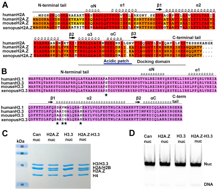
Figure 1.
Sequence conservation of histone variants and nucleosome preparation. (A) Sequence alignment of variant H2A.Z and canonical H2A, showing identical sequences between mouse and human H2A.Z. Conserved amino acids among the three H2A.Z proteins and H2A are highlighted in red. Residues conserved in H2A.Z across all three species are marked in orange, while those conserved only between human and mouse are shown in yellow. Structural elements are indicated above alignment. Docking domain indicated as a dotted line under the alignment. (B) Sequence alignment of the canonical H3.1 and the variant H3.3, showing identical sequences between mouse and human H3.3. Conserved amino acids are in purple. The five amino acid substitutions are marked by stars. (C) Purified histone octamers revealed using 15% Coomassie-stained SDS-PAGE gel. (D) In vitro reconstituted nucleosomes revealed using 3% Native-PAGE.

