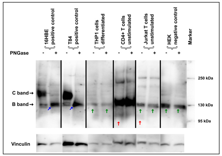
Figure 2.
Glycolytic PNGase digest in immune cells. 16HBE14o- and T84 cells showed a typical shift of both the core-glycosylated CFTR-B and the complex-glycosylated CFTR-C towards the CFTR-A (blue arrow), which constitutes the “naked” protein without any glycosyl side chains, but no such shift was seen for any of the immune cells. In contrast, immunoreactive signals compatible with CFTR-B in size in CD4+ and Jurkat T cells and THP1 cells did not convert to unglycosylated CFTR-A (green arrow). CD4+ and Jurkat T cells displayed non-specific bands at 95 kDa that resolved during PNGase digestion (red arrow).

