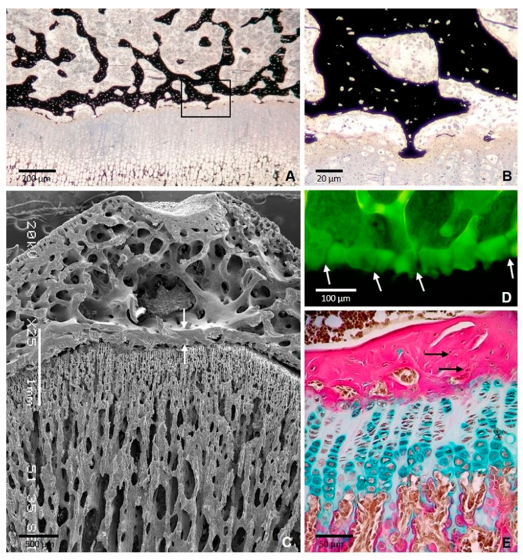
Figure 1.
Formation of the epiphyseal bone plate in the tibial epiphysis of a 35-day-old rat. (A) Section stained with von Kossa showing thin bony extensions bridging the epiphyseal bone plate and the growth plate. The boxed area is shown at a higher magnification in (B) SEM image showing the epiphyseal plate (arrows) and the continuity of the bone plate with the trabecular meshwork of the epiphysis. (C) SEM image showing the epiphyseal plate (arrows) between the trabecular bone of epiphysis and diaphysis. (D) Confocal microscopy image of a 3D projection reconstructed from z-stack images showing the foramina of the plate through which blood vessels passed (arrows). (E) Paraffin section stained with Alcian blue/acid fuchsin showing that the collagen fibers are densely packed but are not arranged in a regular parallel pattern (arrows) [11].

