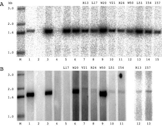
FIG. 4.
Northern analysis to detect dominant-negative inhibition of HDV replication by δAg mutants. (A) Huh7 cells were cotransfected with plasmid pTW148, plasmid pDL444 (which expresses δAg-S), and a construct expressing one of the δAg-S mutants. At 4 days after transfection, total RNA was analyzed and detected as for Fig. 3. Lane M, 5′-labeled single-stranded DNA size markers; lane 1, 2 ng of 1.7-kb HDV cDNA; lane 2, cells transfected with pTW148 alone; lane 3, cells cotransfected with pTW148 and the construct expressing wild-type δAg-S; lanes 4 to 6, triple cotransfections with pTW148, the construct expressing wild-type δAg-S, and a construct expressing δAg-L (lane 4), δAg-S(Δ19–31) (lane 5), or δAg-L(Δ19–31) (lane 6); lanes 7 to 15, alanine mutants as indicated. (B) Experiments essentially as for panel A except that the mutations were expressed on δAg-L rather than δAg-S. Lanes 1 to 4, as in panel A; lanes 5 to 13, alanine mutants as indicated.

