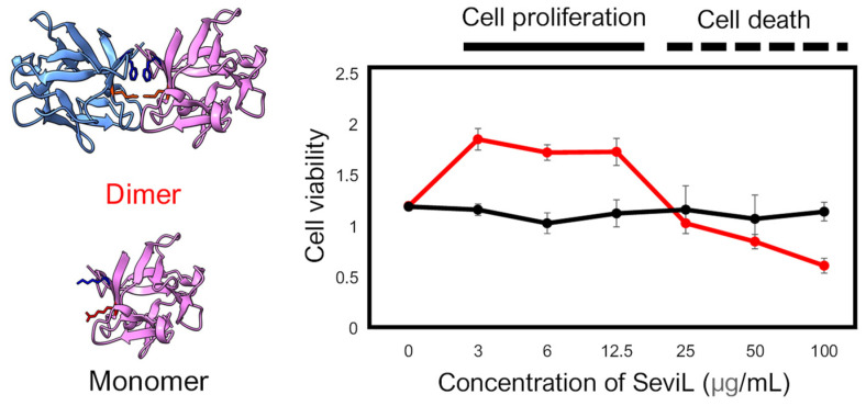
Figure 3.
Comparison of the cellular effect observed in the RAW264.7 cell line between dimeric SeviL (wild type: left upper and right red line) versus monomeric SeviL (mutant: left lower and right black line). Each point represents an average of triplicate measurements. Molecular graphics showing the 3D structures of SeviL were generated by ChimeraX [13].

