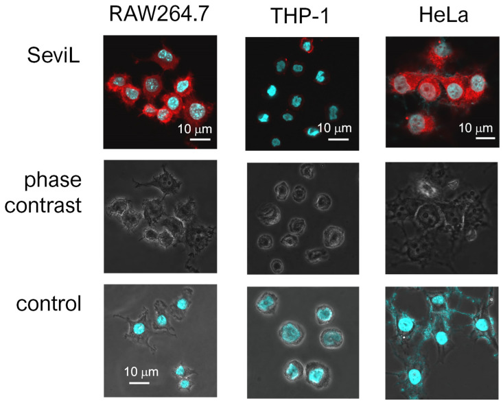
Figure 4.
Different affinities of SeviL for cell membranes. Paraformaldehyde-fixed cells were observed via phase contrast and luminescent scanning microscopy. Staining was performed with fluorescent-labeled SeviL (red) and Hoechst (blue). Scale bar: 10 μm. Controls indicate only Hoechst staining of each cell line.

