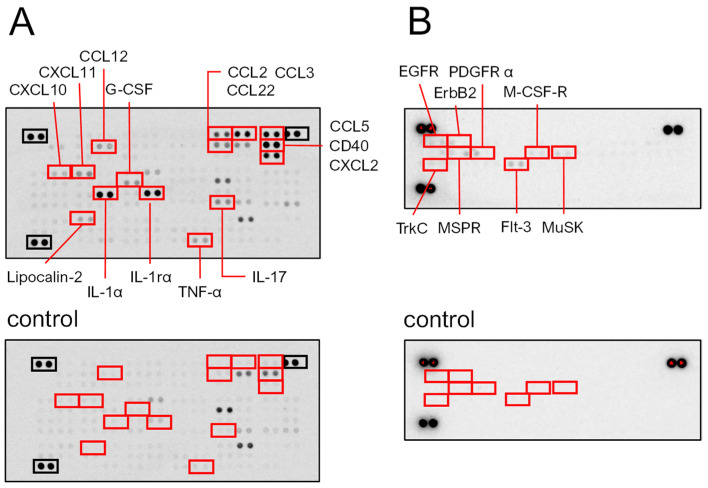
Figure 7.
Macrophage RAW264.7 cells were cultured with or without SeviL (10 μg/mL) for 24 h, and each lysate was applied to a proteome profiler mouse cytokine array (A and A control). The cells were cultured with or without SeviL (10 μg/mL) for 5 min, and each lysate was applied to a receptor tyrosine kinase antibody array (B and B control). The red boxes indicate the proteins whose expression differed significantly between the SeviL-treated and untreated cells in the panel. The black boxes indicate positive controls.

