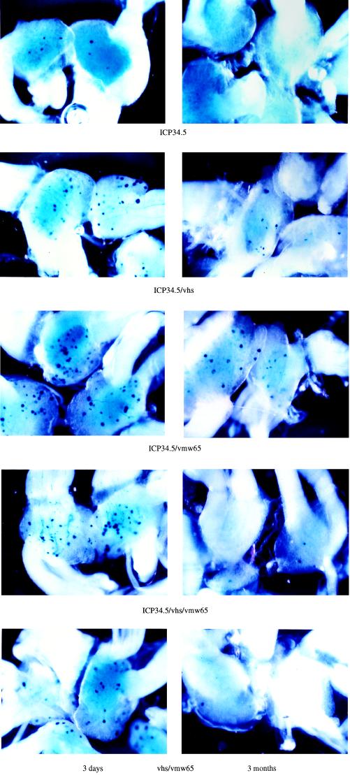
FIG. 4.
Comparative gene delivery to DRGs using various replication-competent HSV vectors. The vectors shown in Fig. 3 were footpad inoculated into 3-week-old BALB/c mice (see Materials and Methods), and DRGs were removed and stained with X-Gal at either 3 days or 3 months postinoculation. L4 and L5 ganglia on the side of injection were removed from pairs of animals. Gene expression levels in L4 and L5 ganglia were in each case found to be similar. Not all DRGs are shown in some panels. In each case the genes inactivated in the respective viruses are indicated.

