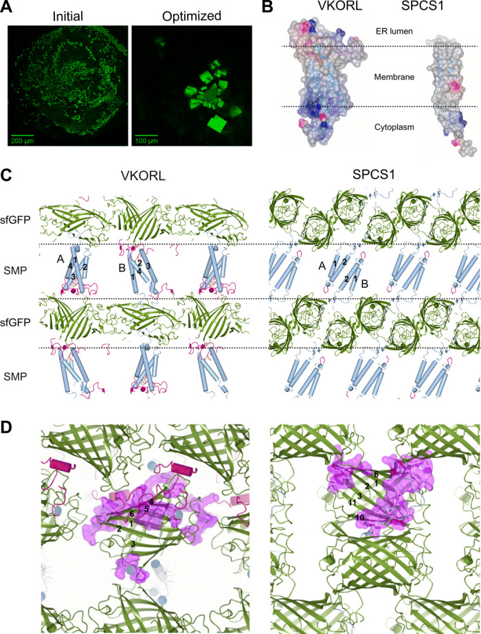
Fig. 3. The sfGFP coupler enables crystallization of small membrane proteins.

VKORL and SPCS1 are shown as examples here; other proteins are shown in fig. S2. (A) Identification of initial crystallization hits (left) of sfGFP-restrained VKORL through fluorescence imaging. The optimized crystals (right) are shown for comparison. (B) Electrostatic surfaces of VKORL and SPCS1 showing their different shapes and surface charges (blue, positive charge; red, negative charge). (C) sfGFP serves as a crystal packing scaffold that is highly adaptable to accommodate various small membrane proteins. Each crystal is packed with alternative layers (dashed lines) of sfGFP (green) and small membrane protein (SMP) molecules. The TMs (numbered) are shown in blue, and extramembrane regions are shown in red. Different membrane protein molecules in the crystals are indicated (A and B). (D) Zoom-in view of versatile crystal packing interactions formed among sfGFP molecules. The large polar surface of sfGFP can accommodate a multitude of crystal contacts (purple surface). The β strands in sfGFP are numbered to illustrate location of the contacts.
