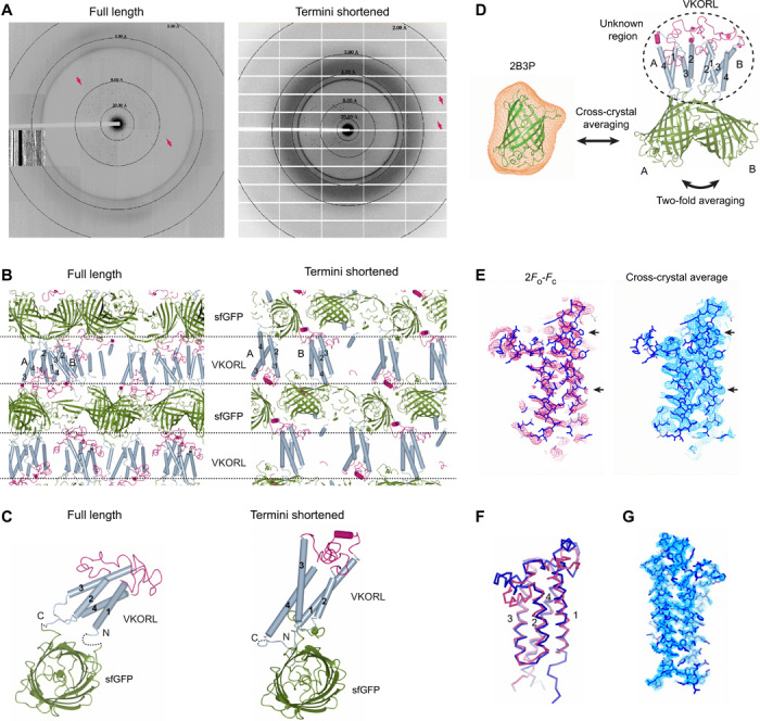
Fig. 4. Restraining-based methods for crystal diffraction and phase improvements.

(A) Shortening the restrained termini improves the crystal diffraction of VKORL. Diffraction patterns are shown for full-length (left) and termini-shortened (right) VKORL restrained by sfGFP. Arrows indicate visible diffraction spots near the diffraction limit. (B) Change of crystal packing interactions. The coloring and annotations are the same as in Fig. 3C. (C) Change of relative orientation between the membrane protein and sfGFP after termini shortening. The dashed lines indicate disordered termini. (D) Improvement of electron density maps through cross-crystal averaging with the coupler protein. The cross-crystal averaging is performed between the sfGFP (2B3P) crystal (averaging mask shown in orange) and the sfGFP-restrained VKORL crystal. In addition, the twofold NCS (molecules A and B) is used. (E) Electron density maps of the VKORL region before and after cross-crystal averaging of the low-resolution data. Arrows indicate regions with improved electron density. (F) Superimposition of the low-resolution (4.3 Å; blue) and high-resolution models (2.4 Å; red) showing that the cross-crystal averaged map allows building of a relatively reliable model at low resolution. (G) Electron density maps of the high-resolution VKORL structure after solvent flattening. All the maps are contoured at 1σ.
