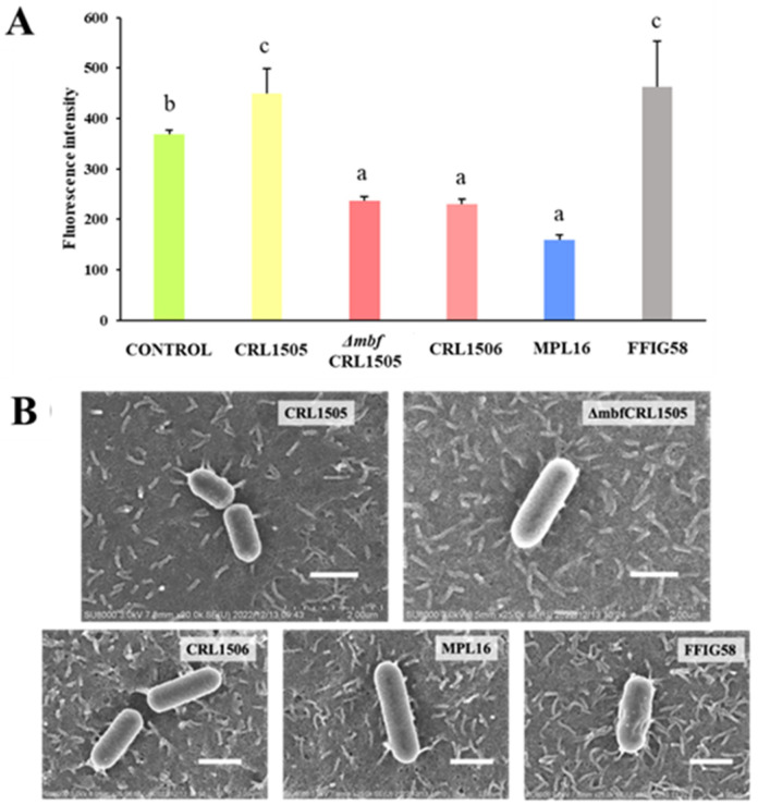
Figure 1.
(A) Adhesion of wild-type Lacticaseibacillus rhamnosus CRL1505 and L. rhamnosus Δmbf CRL1505 to PBE cells. Data from three independent experiments are shown. Different letters indicate statistically significant differences (p ≤ 0.05). (B) Scanning electron microscope (SEM) analysis of wild-type L. rhamnosus CRL1505, L. rhamnosus Δmbf CRL1505, Lactiplantibacillus plantarum CRL1506, L. plantarum MPL16, and Ligilactobacillus salivarius FFIG58 adhering to PBE cells. Scale bar: 1 μm.

