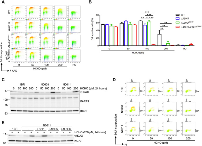
Fig. 2. Formaldehyde is a possible endogenous aldehyde metabolized by both ADH5 and ALDH2.

(A) Formaldehyde treatment inhibits DNA replication in ADH5 and ALDH2 double-deficient cells. 5-ethynyl-2′-deoxyuridine (EdU) incorporation in WT, ∆ADH5, ALDH2E504K, or ∆ADH5 ALDH2E504K double-mutant U2OS cells after formaldehyde treatment is measured by fluorescence-activated cell sorting (FACS) analysis. Cells were incubated with indicated concentration of formaldehyde or 10 mM hydroxyurea (HU) as a positive control for 8 hours followed by EdU incorporation for 1 hour. Then, cells were fixed with 70% ethanol and analyzed by FACS for Alexa Fluor 488–labeled EdU and DNA stained with 7-aminoactinomycin D (7-AAD). Representative FACS images are shown. (B) Quantification of data in (A). Graph shows the percentage of EdU-positive cells. Means (± SD) from three independent experiments are shown. *P < 0.05, **P < 0.01, and ***P < 0.001, one-way ANOVA with Tukey’s multiple comparisons test. (C) Formaldehyde treatment induces DNA damage in cells from AMeDS-affected individuals. Immunoblots showing phospho-Ser139 histone H2AX (γH2AX), a DNA damage marker, and PARP1, an apoptosis marker in normal (1BR) and AMeDS (N0608 and N0611) cells. KU70 is a loading control. (D) EdU incorporation in normal and AMeDS cells after formaldehyde treatment measured by FACS analysis. Cells were incubated with indicated concentration of formaldehyde for 22 hours followed by EdU incorporation for 2 hours. Then, cells were fixed with 70% ethanol and analyzed by FACS for Alexa Fluor 488–labeled EdU and DNA stained with propidium iodide (PI). (E) Formaldehyde-induced DNA damage in AMeDS cells (N0611) is ameliorated with expression of either the wild-type ADH5 or ALDH2 cDNA. Green fluorescent protein as a mock control.
