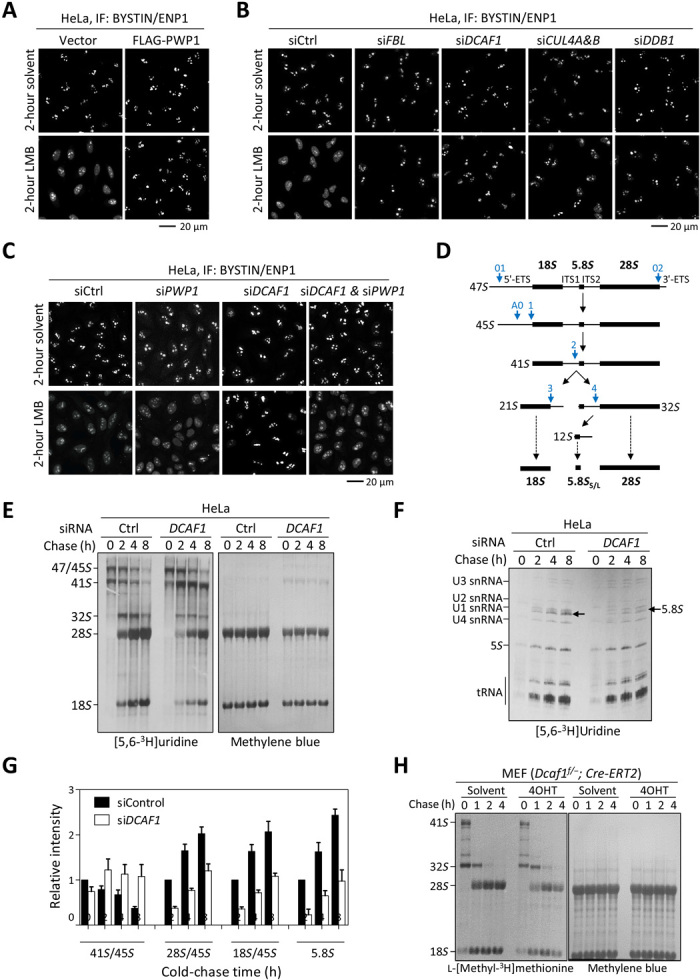
Fig. 5. Loss of function of DCAF1 results in defects in ribosome biogenesis.

(A) HeLa cells stably expressing PWP1 or empty vector were treated with 20 nM LMB or solvent for 2 hours. The subcellular distribution of BYSTIN/ENP1 was determined by IF. (B and C) HeLa cells were transfected with siRNAs targeting indicated genes individually or in combination and then treated with 20 nM LMB or solvent for 2 hours. (D) Diagram illustrating the major steps of mammalian rRNA processing. Blue arrows indicate the major cleavage sites in human cells. (E and F) HeLa cells were transfected with siRNAs targeting DCAF1 or control siRNA and then followed by pulse-chase analysis of the [3H]uridine-labeled rRNAs. The newly synthesized and total rRNAs were determined by autoradiography and methylene blue staining, respectively. (G) The relative intensities of newly synthesized 41S, 28S, and 18S rRNAs compared to 45S precursors at 0 hours in (E) were quantified. The intensities of newly synthesized 5.8S rRNAs in (F) were quantified and normalized against 5S rRNAs. Error bars represent ±SD for duplicate experiments. (H) Dcaf1f/−;Cre-ERT2 MEFs were treated with 200 nM 4OHT or solvent for 3 days, followed by pulse-chase analysis of the l-[methyl-3H]methionine–labeled rRNAs.
