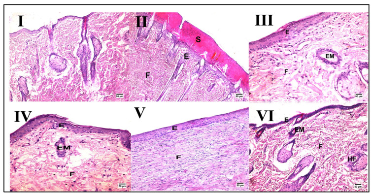
Figure 2.
Representative photomicrographs of histopathological alterations after wound induction (hematoxylin and eosin stain; scale bar 50 μm, 200×). Photomicrographs of skin tissue from the normal control group (I) with the normal histological structure of the skin, the wound injury group (II) showing the formation of a large scab (S) covering some newly formed epidermal layers (E) with fibrous connective tissue formation (F), the PSO/ST-treated group (III) showing migration of epidermal cells (EM) and formation of some layers of epidermis (E) and well-organized fibrous connective tissue (F), the HSO-treated group (IV) presenting migration of epidermal cells (EM) and formation of some layers of epidermis (E), the CSO-treated group (V) showing well-organized fibrous connective tissue (F) and an epidermal layer (E) at the site of the defect, and finally, the ZSO-treated group (VI) showing migration of epidermal cells (EM) and formation of some layers of epidermis (E) with the presence of ill-organized fibrous connective tissue (F).

