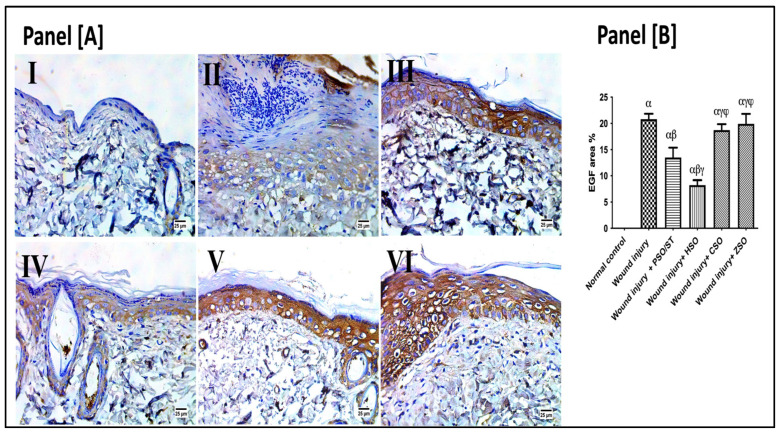
Figure 6.
Representative photomicrographs of the immunohistochemistry expression of epidermal growth factor (EGF) after 14 days of wound induction. Panel [A]: skin sections of the newly formed epidermal layer showing areas stained for EGF expression in the normal skin of healthy rats (I), wound injury group (II), PSO/ST-treated rats (III), rats treated with HSO (IV), CSO (V), and ZSO (VI). Panel [B] represents EGF area% expression (the mean of 10 microscopic fields ± SD) in all treated groups. One-way ANOVA and subsequent multiple comparisons using Tukey’s test as compared with the normal control group (α) and the wound injury (β), wound injury + PSO/ST (γ), and wound injury + HSO (φ), groups [F-value of EGF% = 197.3]. CSO; cantaloupe seed oil; EGF; epidermal growth factor; IHC; immunohistochemistry; HSO; honeydew melon seed oil; PSO; pumpkin seed oil; ST; standard; ZSO; zucchini seed oil.

