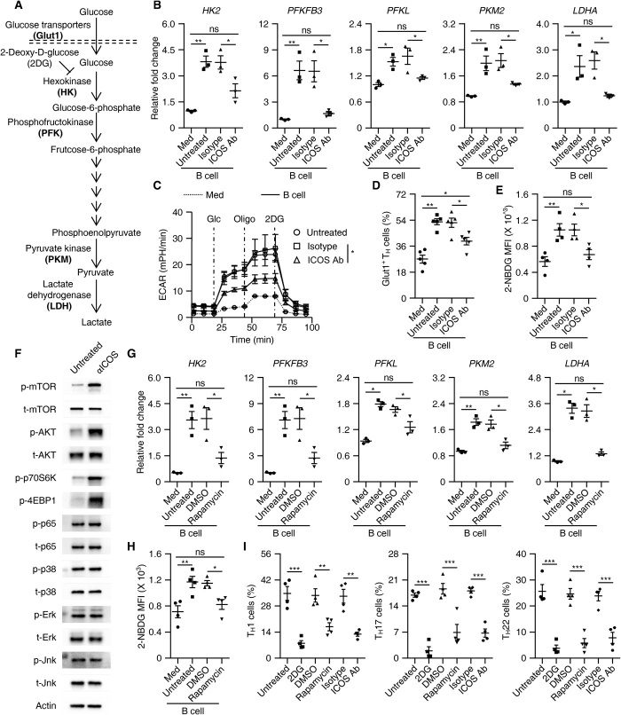
Fig. 5. ICOSL-ICOS axis–elicited glycolysis is involved in T cell polarization by B cells.
(A) Process and key rate-limiting enzymes during glycolysis. (B to E) Purified T cells were cultured in medium or with total B cells in the presence or absence of isotype antibody or ICOS-neutralizing antibody for 7 days, as described in Materials and Methods. Expression of key rate-limiting glycolytic enzymes, ECAR, and capabilities of glucose incorporation (Glut1+ or 2-NBDG intensity) were determined using real-time PCR (B, n = 3), a Seahorse Extracellular Flux XF-24 analyzer (C, n = 3), and FACS (D and E, n = 5 for each), respectively. (F) TCR-primed T cells were left untreated or were stimulated with ICOS agonist antibody for 1 hour. Activation of the indicated signals was detected by immunoblotting (n = 4). (G and H) Using rapamycin to inhibit the mTOR signal impaired B cell–mediated up-regulation of key rate-limiting glycolytic enzymes (G, n = 3) and the incorporation of glucose in TH cells (H, n = 4). DMSO, dimethyl sulfoxide. (I) Using 2DG, rapamycin, and ICOS-neutralizing antibody to inhibit glycolysis, the mTOR signal, and the ICOS signal, respectively, suppressed B cell–meditated inflammatory TH subset differentiation on day 7 (n = 5). Data are representative of three independent experiments (B to I). Data are presented as means ± SEM (B to E and G to I). *P < 0.05, **P < 0.01, and ***P < 0.001 (one-way ANOVA test).

