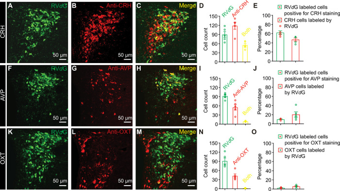
Fig. 1. CRHPVN neurons directly innervate HcrtLH neurons with monosynaptic contacts.

(A to E) Representative slice containing PVN neurons labeled by RVdG (A), antibody staining against CRH (B), merged slice (C), cell counts of RVdG-labeled neurons, CRH-positive neurons and both-positive neurons (D), and percentages of RVdG-labeled neurons positive for CRH staining and CRH neurons labeled by RVdG (E). (F to J) Representative slice containing PVN neurons labeled by RVdG (F), antibody staining against AVP (G), merged slice (H), cell counts of RVdG-labeled neurons, AVP-positive neurons and both-positive neurons (I), and percentages of RVdG-labeled neurons positive for AVP staining and AVP neurons labeled by RVdG (J). (K to O) Representative slice containing PVN neurons labeled by RVdG (K), antibody staining against OXT (L), merged slice (M), cell counts of RVdG-labeled neurons, OXT-positive neurons and both-positive neurons (N), and percentages of RVdG-labeled neurons positive for OXT staining and OXT neurons labeled by RVdG (O) (n = 5 mice).
