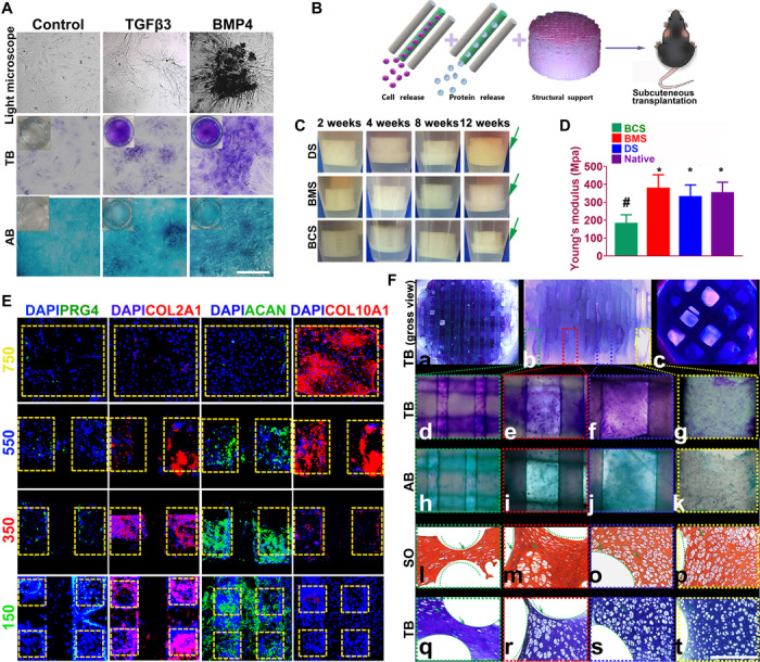
Fig. 4. Dual-factor releasing induced cartilaginous matrix formation in 3D bioprinted gradient-structured scaffolds.

(A) Chondrogenic differentiation of condensed rMSCs with toluidine blue (TB) and alcian blue (AB) staining. (B) Scaffolds were transplanted subcutaneously for 12 weeks. (C) To validate the cartilage-generating capability, scaffolds were incubated and observed for 12 weeks in vitro, indicating better cartilage-generating potential for the physically gradient protein-releasing scaffold (movie S2). (D) Young’s modulus of the scaffolds compared with native cartilage after 12 weeks. Data are presented as averages ± SD (n = 6). *P < 0.05 between the NG-750 group and other groups; #P < 0.05 between the native cartilage group and other groups. (E) In the generated cartilage tissues, spatiotemporally released dual-factors induced zone-specific expression of PRG4, aggrecan, and collagens II and X and showed resemblance with native joint cartilage. (F) (a to c) Toluidine blue staining of the 3D printed cartilage constructs (a, top view; b, side view; c, bottom view) after culture in chondrogenic medium for 6 weeks in vitro. (d to g) Toluidine blue and (h to k) alcian blue staining was applied for each layer of the gradient scaffold. (l to p) Safranin O (SO) and (q to t) toluidine blue staining of cartilage tissue between PCL fibers (green curved line) in different layers of the 3D printed cartilage constructs after subcutaneous implantation. Photo credit: Ye Sun, First Affiliated Hospital of Nanjing Medical University.
