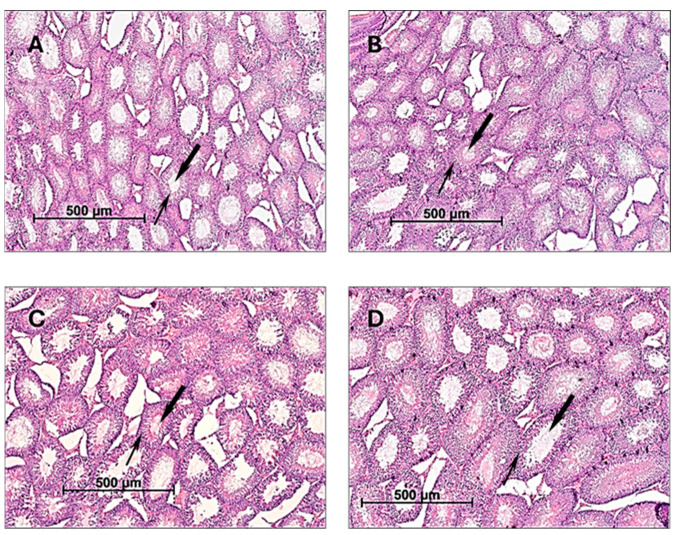
Figure 1.
The morphological structure of testes of male rats. Male rats were orally treated for 60 days with (A)—H2O (control); (B)—CE 100 mg/kg bw/day; (C)—500 mg/kg bw/day; and (D)—1000 mg/kg bw/day. Thin arrow—seminiferous spermatogenic epithelium of the testes; bold arrow—lumens of the seminiferous tubule. Staining hematoxylin/eosin; magnification ×50.

