Figure 3.
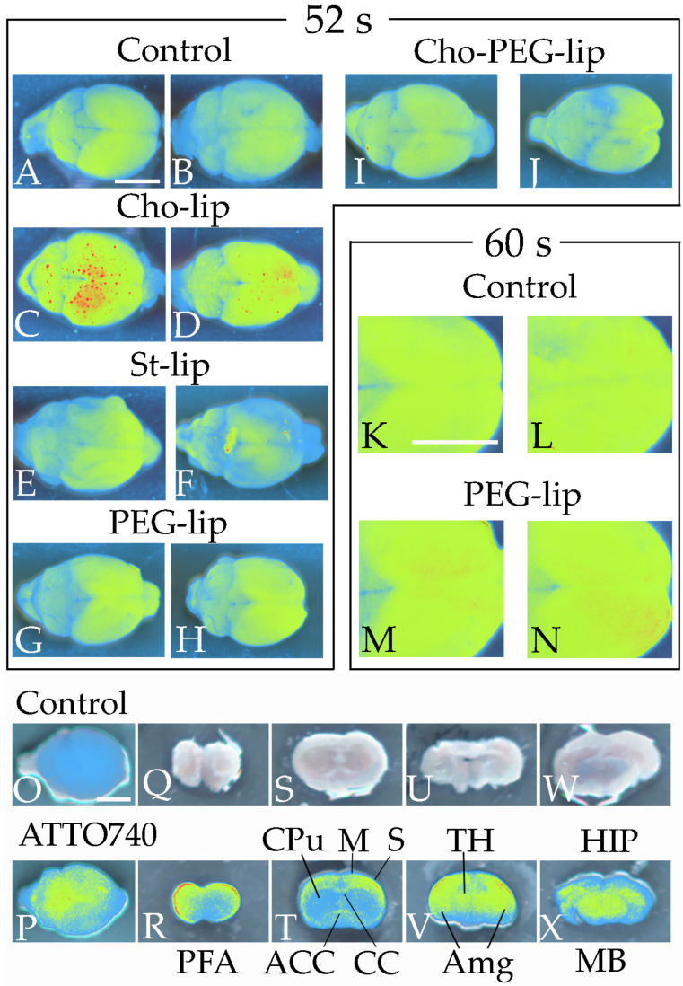
DiIC18 and ATTO 740 DOPE, incorporated into liposomes, accumulate in specific brain areas as revealed by in vivo fluorescence imaging. Fluorescence images of the dorsal side of the brain were acquired 3 h after control or fluorescent liposome injection (A–N). Compared with mice injected with control liposomes (A,B), strong fluorescence (red) was observed in the cerebrum and midbrain of mice injected with Cho-lip (C,D) at 52 s of exposure, but not in mice injected with St-lip (E,F), PEG-lip (G,H), or Cho-PEG-lip (I,J). At 60 s of exposure (K–N), fluorescence in the cerebrum was stronger in mice who received liposomes containing PEG than in mice injected with control liposomes. Similar to DiIC18, fluorescence was detected in the cerebrum and midbrain after the injection of liposomes with ATTO 740 DOPE (O,P). In the coronal brain slices (Q–X), ATTO 740 DOPE accumulated in the prefrontal area (PFA), motor cortex (M), somatosensory cortex (S), thalamus (TH), amygdala (Amg), hippocampus (HIP), midbrain (MB), anterior commissure (ACC), and corpus callosum (CC), but not in the caudate putamen (CPu). Scale bar: 5 mm.

