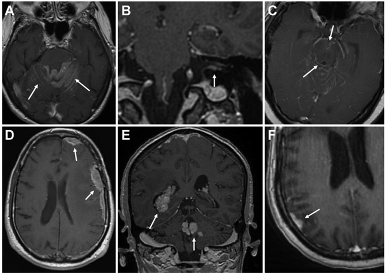
FIG. 1.
Imaging features of LMD subtypes. A–C: Axial and coronal T1-weighted postcontrast MR images demonstrating cLMD (arrows) involving abnormal “sugarcoating” along cerebellar folia (A), along cranial nerves and within the internal acoustic canal (B), and along the brainstem (C), in addition to other regions. D–F: Axial and coronal T1-weighted postcontrast MR images demonstrating nLMD (arrows) involving nodular, dural-based (D and F), or ependymal (E) lesions.

