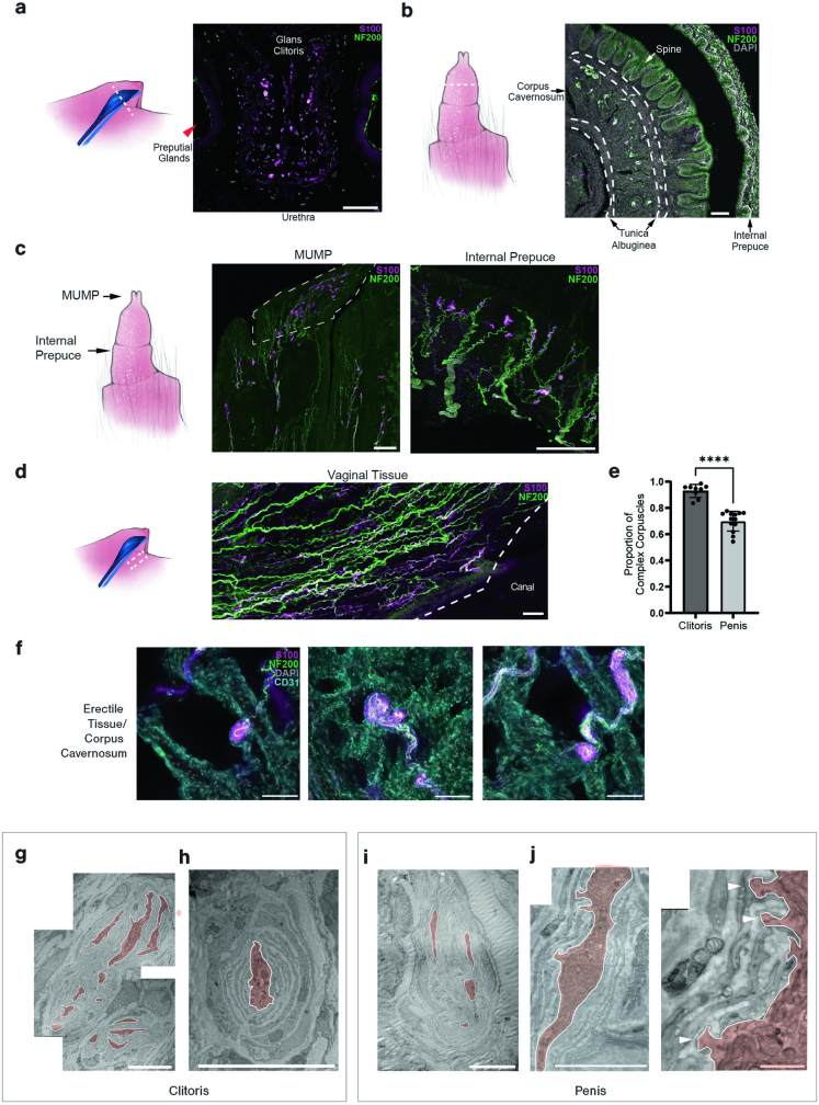
Extended Data Fig. 1. Distribution and morphological features of Krause corpuscles.
a, Immunostaining of a coronal section through the clitoris and surrounding tissue (left dashed line) for S100 and NF200. The clitoris, urethra, and preputial glands are annotated. b, Immunostaining of a coronal section through the penis as in a. The internal prepuce, penile spines of the glans penis, corpus cavernosum, and surrounding tunica albuginea are annotated. c, Corpuscles within the male urogenital mating protuberance (MUMP) and internal prepuce are visualized as in a. d, Immunostaining of a section of vaginal tissue, stained as in a, isolated from the dashed area, shows an absence of Krause corpuscles. e, Quantification of the percent of complex Krause corpuscles among the total corpuscles observed in a section of genital tissue (9 sections from 4 females and 13 sections from 4 males). ****p < 0.0001, unpaired t-test. f, Immunostained sections showing Krause corpuscles within the corpus cavernosum of erect penis tissue for S100, NF200, and CD31. g, Representative transmission electron micrograph of a multi-lobulated complex Krause corpuscle within the clitoris. h, Example electron micrograph of a simple Krause corpuscle with a single lamellated axon profile within the clitoris. i, Example electron micrograph of a complex Krause corpuscle within the penis. j, Example electron micrograph of a Krause corpuscle (left) magnified to visualize axonal protrusions (right, white arrowheads). All axon profiles in g-j were manually outlined and filled (light red). Scale bar is 100 µm in a-d, 50 µm in f, 10 µm in g-i, 5 µm in j (left), 1 µm in j (right). Error bars, s.d. The diagrams in a–d were created by G. Park.

