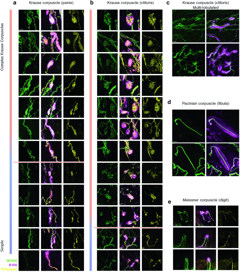
Extended Data Fig. 2. The diversity of Krause corpuscle morphologies and comparison with Meissner and Pacinian corpuscles.
The range of Krause corpuscle morphologies found in the penis a and clitoris b, from complex (top) to simple (bottom). The red line depicts the distinction between complex (above the line) and simple (below the line) Krause corpuscles for quantification in Fig. 1e. c, Examples of multi-lobulated Krause corpuscles observed in the clitoris. d, Examples of Pacinian corpuscles located in the periosteum of the fibula. e, Examples of Meissner corpuscles located in the glabrous dermal papilla of a forepaw digit tip. The scale bar is 20 µm, and all images are of the same scale. Images in a, b and e are stained for NF200 (green), S100 (magenta), and tdTomato for TrkB axons (yellow) in TrkBCreER; AvilFlpO; R26FSF-LSL-Tdtomato mice. Images in c and d are stained for NF200 (green) and S100 (magenta).

