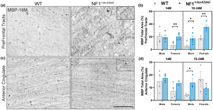
FIGURE 1.

NF1 +/ex42del miniswine exhibit increased MBP+ mature oligodendrocytes in prefrontal tracts and anterior cingulate. (a) Representative images of MBP+ oligodendrocytes in the prefrontal tracts of wild‐type and NF1 +/ex42del miniswine at 18 months of age. (b) MBP+ analysis in prefrontal tracts indicating significant differences between NF1 +/ex42del miniswine and wild‐type counterparts at 14 and 18–24 months of age. (c) Representative images of MBP+ oligodendrocytes in the anterior cingulate of wild‐type and NF1 +/ex42del miniswine at 18 months of age. (d) MBP+ analysis in the anterior cingulate indicates significant differences between male NF1 +/ex42del and wild‐type counterparts at 18 months. Unpaired t‐tests. n = 3–4/group, *p < 0.05, **p < 0.01. Mean ± SEM. Scale bar = 200 μm.
