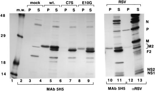
FIG. 4.
Analysis of the effects of mutations in the Cys3-His1 motif of M2-1 on phosphorylation. HEp-2 cells infected with vTF7-3 were mock transfected (mock lanes 2 and 3) or transfected with a plasmid bearing a gene encoding the wild-type or a mutant M2-1 protein as indicated (lanes 4 to 9), or HEp-2 cells were infected with RS virus (RSV lanes 10 to 13). Cells were exposed to [35S]methionine and [35S]cysteine (S lanes 3, 5, 7, 9, 11, and 13) or [33P]inorganic phosphate (P lanes 2, 4, 6, 8, 10, and 12) for 2 h at 16 h posttransfection or 20 h postinfection. Labeled proteins were immunoprecipitated using M2-1-specific MAb 5H5 (lanes 2 to 11) or anti-RS virus polyclonal serum (αRSV lanes 12 and 13) and analyzed by SDS-PAGE in 11% polyacrylamide gels followed by fluorography. Positions of RS virus proteins are shown (lane 13). The exposure time of lanes 10 to 13 was two times that of lanes 1 to 9. m.w., molecular weight markers (sizes in thousands are noted at the left); wt., wild type.

