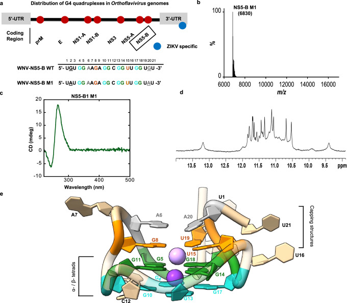
Fig. 1. Solution state characterization of NS5-B M1.
a Location and sequence of the NS5-B in an Orthoflavivirus genome (top). Wild-type (WT) and mutated (M1) NS5-B sequences (bottom). Bases making up the α-tetrad are in teal, the β-tetrad are in green, the γ-triad are in orange, and the dyad in gray. Sequence variation is indicated by underlining. b Mass spectrometry confirms that NS5-B M1 is a monomer with a mass of ~7 kDa. c The CD spectrum is characteristic of parallel topology with a maximum at ~264 nm and a minimum at ~245 nm. d The 1D 1H imino proton spectrum of NS5-B M1 at 298 K in the presence of K+ ions exhibits characteristic quadruplex signals at 10–12 ppm and is consistent with the spectrum of the WT sequence (Fig. S1a). e A view of the 1.97 Å x-ray crystal structure of the G2A/G20A mutated NS5-B M1 West Nile Virus quadruplex. Additional views are provided in Movie S1. Source data for (b, c) are provided as a Source Data file.

