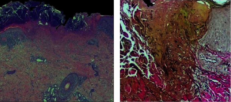
Figure 2.

Histopathological images. (a) A broad epidermal ulceration with extensive scale crust. (b) Vertically oriented degenerated and eosinophilic collagen fibers together with black elastic fibers are extruded through the epidermal breach. ((a) Hematoxylin-eosin stain, original magnification: 20x; (b) Verhoef–Van Gieson stain, original magnification: 100x).
