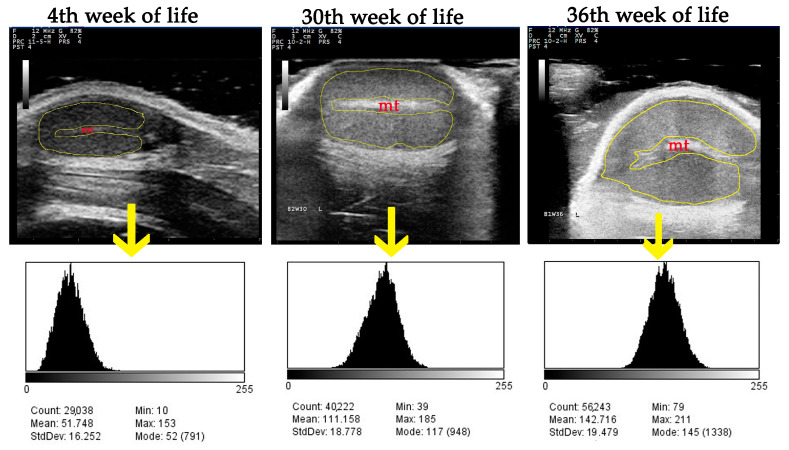
Figure 3.
The region-area technique of grayscale analysis: the entire image of testicular parenchyma is outlined (yellow line), excluding mediastinum testis (mt), and corresponding histograms are created for the calculation of mean pixel grayscale intensity (numerical pixel values) and pixel heterogeneity (pixel values standard deviations).

