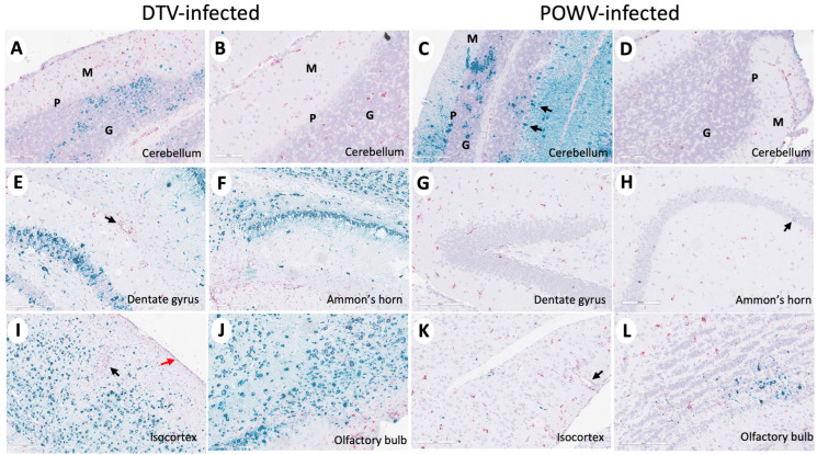
Figure 5.
Divergent patterns of infection in DTV- and POWV-infected mouse brains. Brains were harvested at the time of euthanasia. Representative images of DTV-infected (A,B,E,F,I,J) and POWV-infected (C,D,G,H,K,L) brains stained for viral RNA (teal) and CD11b+ cells (pink). (A–D) Molecular (green M), Purkinje (green P), and granular (green G) cell layers of the cerebellum and (C) POWV-infected Purkinje cells (black arrow). (E) CD11b positive cells (black arrow) around a vessel in the hippocampal region of a DTV-infected mouse brain. (H) Minimal infected cells (black arrow) in the hippocampal region of a POWV-infected mouse brain. (I) CD11b positive cells around a vessel (black arrow) and in the leptomeninges (red arrow) of the isocortex of a DTV-infected mouse brain. (K) CD11b positive cells in the perivascular (Virchow–Robin) space (black arrow) of a POWV-infected mouse brain.

