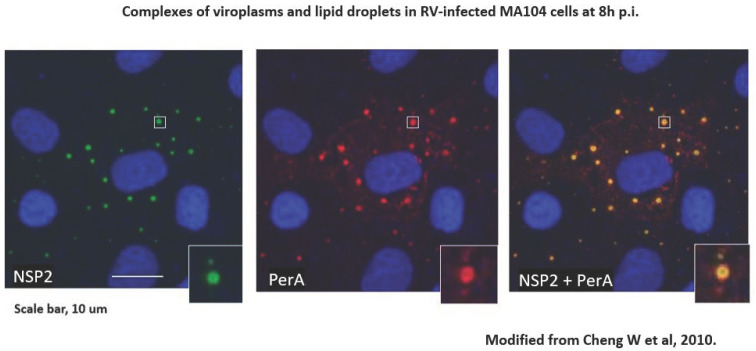
Figure 1.
RV-infected cells grown on coverslips were processed for confocal microscopy (CM) as described [20]. Cells were blocked with 1% BSA–0.1% triton X-100. Primary antibodies against perilipin A (rabbit polyclonal) were from Abcam, and those against NSP2 (mouse monoclonal) were a kind gift of Dr Oscar Burrone, ICGEB, Trieste IT. Secondary antibodies were goat anti-rabbit IgG conjugated with Alexafluor 633 and goat anti-mouse IgG conjugated with Alexafluor 488, both from Invitrogen. Staining with secondary antibodies was carried out in the presence of 2 µg/mL of Hoechst 33,342 (from Sigma). Coverslips were mounted on glass slides with Prolong bold Antifade mounting medium (from Molecular Probes) and observed by CM using a Leica DM Libre TCS SP instrument. An individual viroplasm–LD complex is magnified in the inserts. From [17].

