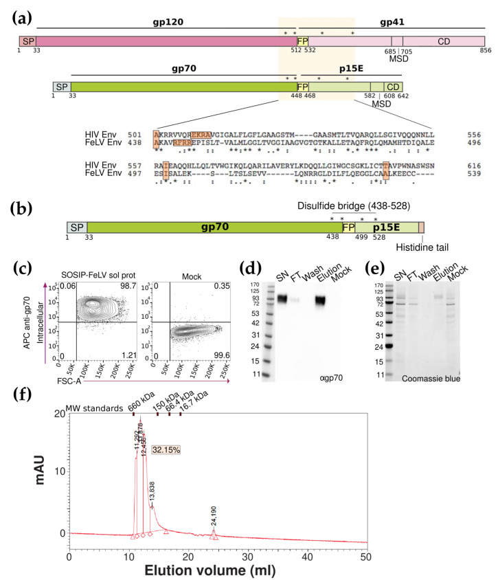
Figure 1.
Generation of a soluble stable Env trimer. (a) Schematic representation of HIV and FeLV Env glycoproteins, mutations of SOSIP strategy are indicated by asterisks. Alignment between HIV and FeLV Env glycoproteins. (b) The schematic representation of the SOSIP FeLV soluble protein. Signal peptide (SP), mutations (indicated by asterisks), disulfide bridge, and histidine tail are denoted. (c) Representative flow cytometry panels for the total expression of the SOSIP-FeLV soluble proteins detected with an anti-gp70 antibody. (d) The Western blot analysis of the different fractions of the SOSIP-FeLV soluble protein purification developed with an anti-gp70 antibody. (e) The Coomassie blue analysis of the different fractions of the SOSIP-FeLV soluble protein purification. (f) Chromatogram using the SE-HPLC of the SOSIP-FeLV soluble protein.

