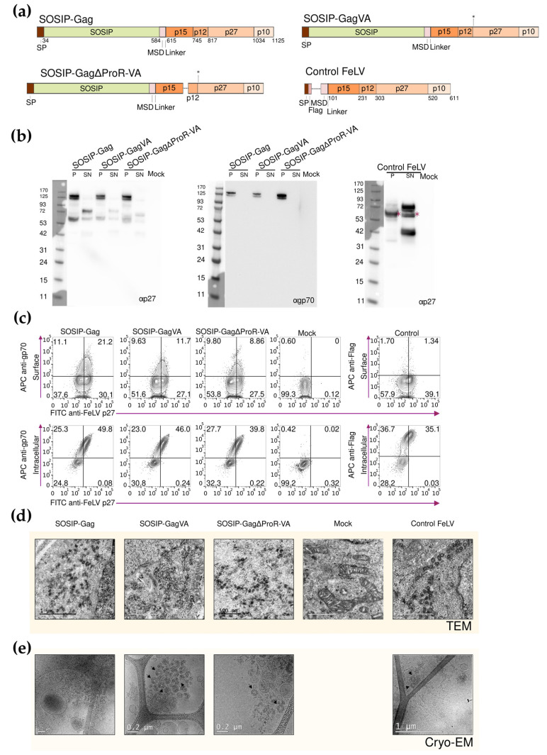
Figure 2.
Design and characterization of the SOSIP-FeLV Gag fusion proteins. (a) The schematic representation of each fusion protein, asterisks indicate location of the VA mutations in FeLV Gag. (b) Western blot was developed with anti-p27 or anti-gp70 antibodies to analyze the expression of the SOSIP-FeLV Gag fusion proteins in the Expi293F cells. Asterisks show estimated molecular weight. (c) Representative flow cytometry panels for the extracellular and intracellular detection of the fusion proteins using anti-gp70, anti-Flag, and anti-p27 antibodies. (d) TEM images were obtained from the Expi293F cells expressing the indicated FeLV Gag-based VLPs. (e) Cryo-EM images were obtained using the purified FeLV Gag-based VLPs.

