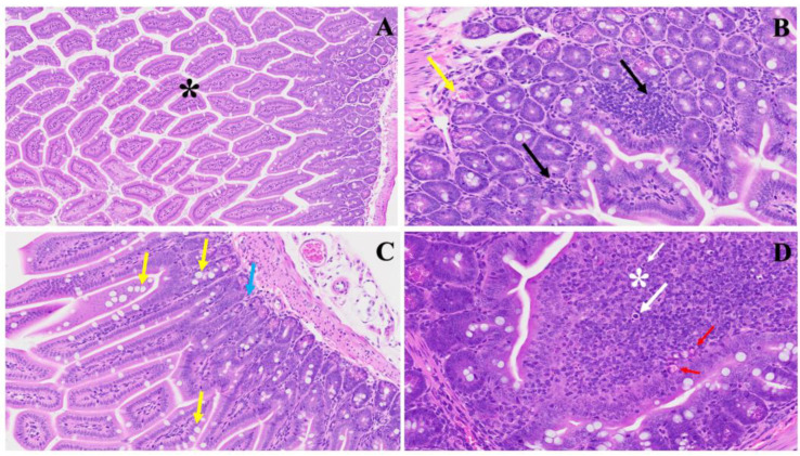
Figure 1.
This illustrates the histopathological changes observed in acute extensive intestinal inflammation in the MHV-1 model in small and large intestines. In Panel (A), the typical architecture of the large intestine is depicted, with intact villi highlighted by a star. Panel (B) demonstrates colonic mucosal inflammation and lymphoid hyperplasia, indicated by black arrows, accompanied by microthrombi marked by yellow arrows. In Panel (C), hyperplasia of goblet cells scattered throughout the colonic mucosa is shown (yellow arrows), along with various inflammatory cells within the lamina propria (blue arrow). Panel (D) reveals diffuse proliferation of lymphoid tissue (white star), microbleeding (red arrow), and melanocytosis (white arrows) in the colonic mucosa. These findings provide insight into the pathological features associated with acute significant intestinal changes in the MHV-1 model. (H&E, original magnification 66× (A–D)). MHV-1 infection alone (n = 16), healthy control (n = 7), and SPK-treated mice (n = 5).

