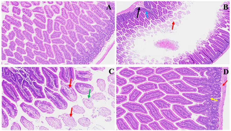
Figure 2.
This shows acute small and large intestinal changes induced by MHV-1 infection. In Panel (A), a histological section displays the typical regular colonic layers. Panel (B) exhibits early manifestations of disease, with evident mucosal layer sloughing (red arrow), concomitant inflammatory alterations in the muscularis mucosa layer (black arrow), and overall inflammation (blue arrow). Panel (C) depicts diverse stages of villus degeneration (red arrows) alongside the presence of crypt apoptotic bodies (green arrow). In Panel (D), regenerative responses during the acute phase of SPK treatment are evident, including the reconstruction of the muscularis mucosa layer (red arrow), normalization of goblet cells (white arrow), restoration of crypt and lamina propria anatomy (yellow arrow), and mitigation of inflammatory changes. (H&E, original magnification 66× (A–D)). MHV-1 infection alone (n = 16), healthy control (n = 7), and SPK-treated mice (n = 5).

