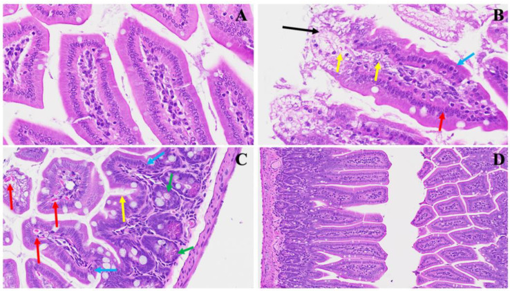
Figure 3.
This portrays the acute small intestinal changes ensuing from MHV-1 infection. In Panel (A), a depiction of normal crypt morphology with intact goblet cells is observed. Panel (B) showcases several mitotic figures within the villi (yellow arrows) alongside sloughing villi (black arrow) and the destruction of enterocytes, representing the simple columnar epithelium (red arrow) accompanied by edema surrounding enteroendocrine cells (blue arrow). Panel (C) further elucidates the presence of edema surrounding enteroendocrine cells (blue arrow), dying Paneth cells (green arrow), increased mucus secretion (yellow arrow), and invasion of red blood cells (red arrows). Panel (D) demonstrates the restoration of regular histopathological changes following SPK administration characterized by reduced sloughing and normalization of goblet cells. These observations offer insights into the dynamic alterations occurring in the small intestine during MHV-1 infection and hint at the potential therapeutic benefits of SPK treatment. (H&E, original magnification 66× (A–C) and 22× (D)). MHV-1 infection alone (n = 16), healthy control (n = 7), and SPK-treated mice (n = 5).

