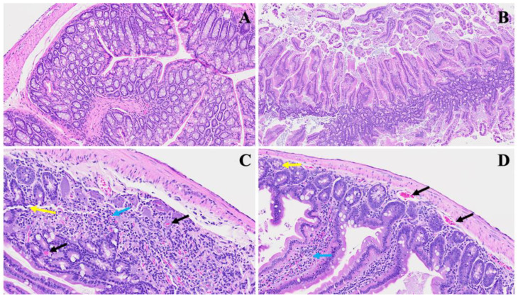
Figure 4.
Representation of acute small bowel damage induced by MHV-1 infection. Panel (A) demonstrates normal small bowel histology, while Panel (B) depicts severe inflammatory changes penetrating all layers of the small intestine, including papillary necrosis, apoptosis, and an inflamed lamina propria. Panel (C) highlights the infiltration of immune cells, with neutrophils (yellow arrow) and lymphocytes (blue arrow) evident alongside observable microthrombi (black arrows). Panel (D) further illustrates microthrombi (black arrows), dying crypts (yellow arrows), and pronounced villi inflammation (blue arrows). (H&E, original magnification 22× (A–D)). MHV-1 infection alone (n = 16), healthy control (n = 7), and SPK-treated mice (n = 5).

