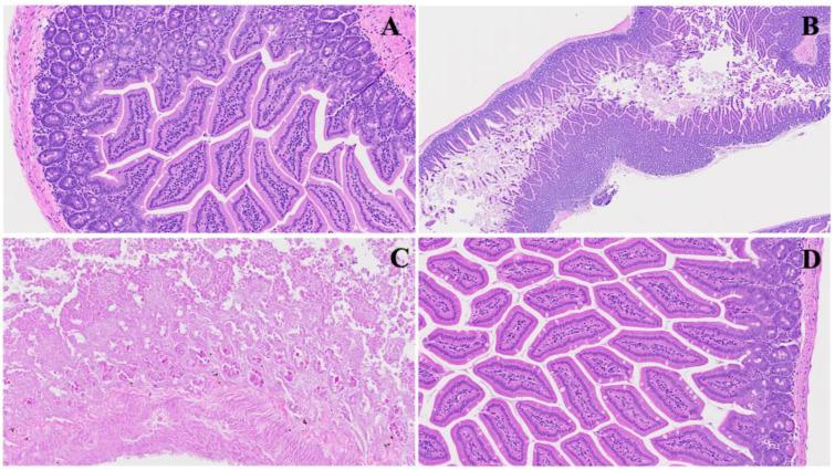
Figure 6.
Acute ileum changes post MHV-1 infection. Panel (A) illustrates the histology of a normal ileum. In Panel (B), acute severe hyperplasia of crypts is observed, accompanied by various stages of inflammatory changes. Panel (C) showcases the massive destruction of one part of the ileum, characterized by the blunting of crypts and villi and the loss of brush borders. Panel (D) depicts the restoration of tissue architecture following SPK treatment. (H&E, original magnification 66× (A,D) and 22× (B,C)). (MHV-1 infection alone (n = 16), healthy control (n = 7), and SPK-treated mice (n = 5)).

