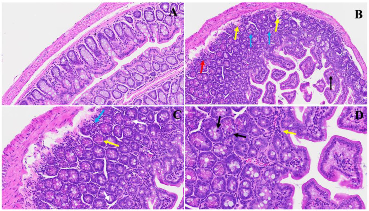
Figure 7.
This illustrates the duodenum intestinal changes associated with long COVID. In Panel (A), a depiction of standard small intestinal architecture is shown. Panel (B) highlights alterations observed in long COVID, including nests of erythrocytosis indicated by yellow arrows, diffused inflammation marked by a red arrow, lymphocyte invasion denoted by blue arrows, along with an increased number of goblet cells, various apoptotic bodies, congestion, and thrombosis indicated by a black arrow. Panel (C) exhibits severe inflammatory cellular invasion (blue arrow), accompanied by an increase in Paneth cells (yellow arrows) and the presence of neutrophils (white arrow). Panel (D) demonstrates various inflammatory cell infiltrates (yellow arrow) alongside erythrocytosis, with evident apoptotic bodies (black arrows) and an increase in Paneth cells. (H&E, original magnification 66× (A–D)). (4 MHV-1 infection, 4 healthy control, 4 SPK treated group).

