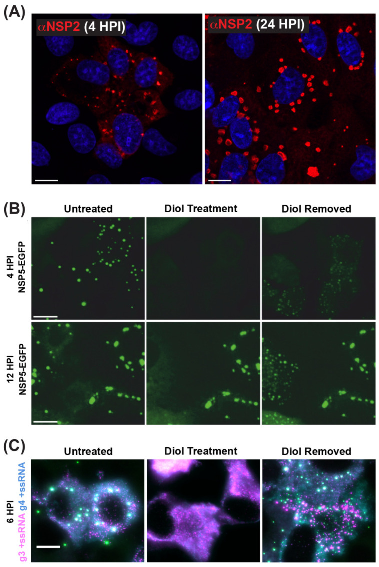
Figure 6.
Viroplasms are Cytoplasmic Biomolecular Condensates Formed via LLPS. (A) Fixed immunofluorescence images of SA11-infected MA104 cells at 4 and 24 h post-infection (HPI). Viroplasms were stained with a polyclonal antibody against NSP2 (αNSP2; red), and cell nuclei were stained with Hoechst (blue). Scale bar = 10 μm. Images adapted from reference [70] with permission. (B) Viroplasms formed in MA104 cells stably expressing NSP5-EGFP and infected with strain SA11. Numerous small viroplasms can be dissolved when low concentrations of aliphatic diols (4.7% propylene glycol or 4% 1.6-hexane diol) are applied directly to the cell culture medium at 4 h post-infection (HPI). Removal of diols from the medium results in reassembly of multiple smaller granules dispersed in the cytosol (right panel). At 12 HPI, viroplasms become larger and less regular in shape. These larger viroplasms become resistant to the application of aliphatic diols. Scale bar = 50 µm. Images adapted from [71] with permission. Copyright Creative Commons Attribution License (https://creativecommons.org/licenses/by/4.0/). (C) RNA FISH imaging of gene segment 3 +ssRNA (magenta, g3 +ssRNA) and gene segment 4 +ssRNAs (cyan, g4 +ssRNA) in SA11-infected NSP5-EGFP-expressing MA104 cells fixed at 6 HPI. Viroplasms were treated with 4.7% (v/v) propylene glycol (middle) at 4 HPI, releasing +ssRNAs into the cytoplasm. These granules reformed after replacing the propylene glycol-containing cell culture medium, resulting in the rapid re-localization of g3 +ssRNA (magenta) and g4 +ssRNA (cyan) transcripts are detected via smFISH, and colocalizing RNAs (white). Scale bar = 10 µm. Images adapted from [72] with permission. Copyright Creative Commons Attribution License (https://creativecommons.org/licenses/by/4.0/).

