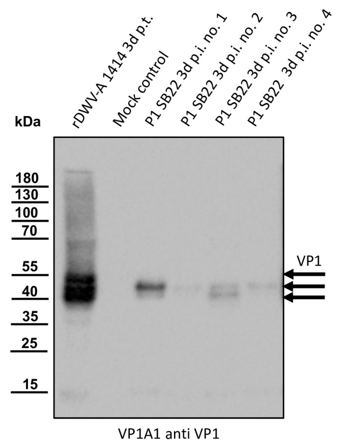
Figure 2.
Western blot analyses of bee pupae infected with the isolate Austria-SB22. As controls, honey bee pupae were transfected with the in vitro-transcribed RNA of DWV strain 1414 (rDWV-A 1414 3d p.t.) or mock-transfected with PBS. Four pupae were injected with 1 µL of a DWV-B-positive bee lysate (P1 SB22 3d p.i. no. 1–4). After a three-day incubation period, the pupae were harvested and homogenized, and the total bee protein was resolved via SDS-PAGE. A typical VP1 pattern with bands at 47, 42, and 39 kDa appeared in the rDWV-A-transfected pupa. The mock-transfected pupa showed no signal, indicating no background infections in our experimental animal. All pupae inoculated with the original sample material of SB22 showed a protein band at 42 kDa. Pupa no. 1 displayed a strong signal, while signals in pupa nos. 2–4 were relatively weak. Pupa nos. 1 and 3 showed an additional signal at 39 kDa. Arrows indicate the protein bands of the characterized target proteins of the antibody used. The bands of a pre-stained molecular weight marker are indicated on the left.

