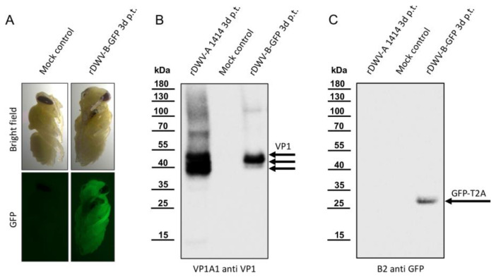
Figure 5.
The employment of a GFP reporter in rDWV-B. (A) The fluorescence of rDWV-B-GFP-transfected bee pupae. The GFP fluorescence emanating from the rDWV-B-GFP-transfected pupa was remarkably intense and already easily visible through filter eyeglasses. Conversely, only negligible background fluorescence was detected in the mock-transfected pupa. (B) VP1 expression of rDWV-B-GFP. Pupae were transfected with either rDWV-A or rDWV-B-GFP or injected with PBS (Mock control) and harvested three days post-transfection. Whole-bee lysates were prepared and subjected to SDS-PAGE, followed by blotting and VP1 analysis. Typical VP1 bands were evident in the positive control (rDWV-A 1414), and a strong VP1 signal was observed in the rDWV-B-GFP-transfected pupa, with main bands appearing at 42 kDa and 39 kDa. No signals were detected in the mock-infected pupa. (C) Processing of the GFP-T2A reporter. Another Western blot was conducted using B2 anti-GFP. Only the rDWV-B-GFP-transfected bee exhibited a signal corresponding to the calculated weight of GFP-T2A at an apparent molecular weight of 29 kDa. The absence of additional bands suggests the effective separation of GFP and the leader protein by the T2A peptide.

