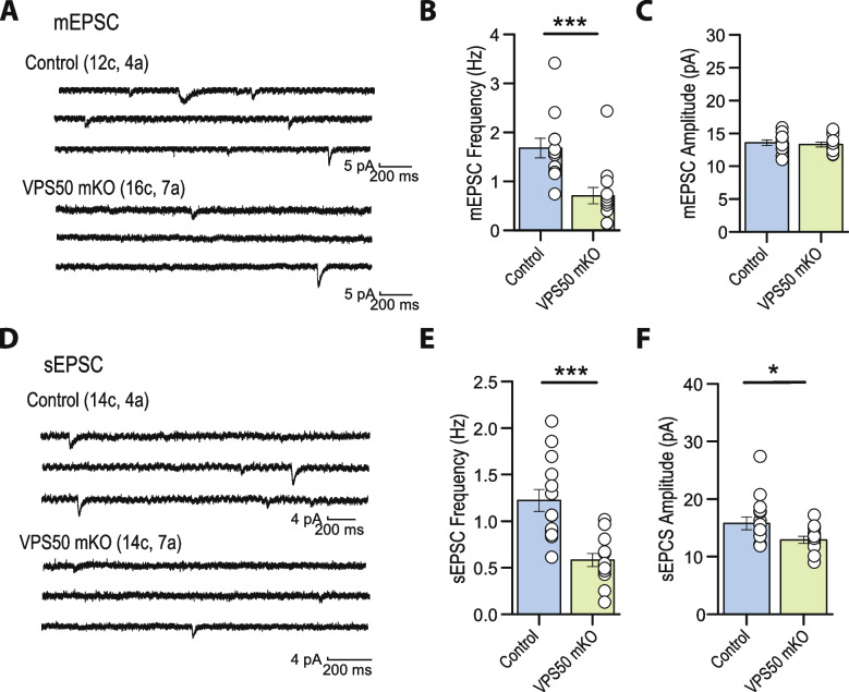
Fig. 5.
Hippocampal spontaneous excitatory synaptic activity is impaired in VPS50 mKO mouse. A Representative traces of mEPSCs recorded from CA1 pyramidal neurons in acute hippocampal slices of control and VPS50 mKO animals. B, C Quantification of the frequency (B) and amplitude (C) of mEPSCs. D Representative traces of sEPSCs in hippocampal slices of control and VPS50 mKO animals. E, F Quantification of the frequency (E) and amplitude (F) of mEPSCs. In each representative trace (A–D), the numbers of cells (c) and animals (a) are indicated in parentheses. Two-samples Student t-test was used for statistical analysis; *p < 0.05, ***p < 0.001. Summary data consist of mean ± SEM

