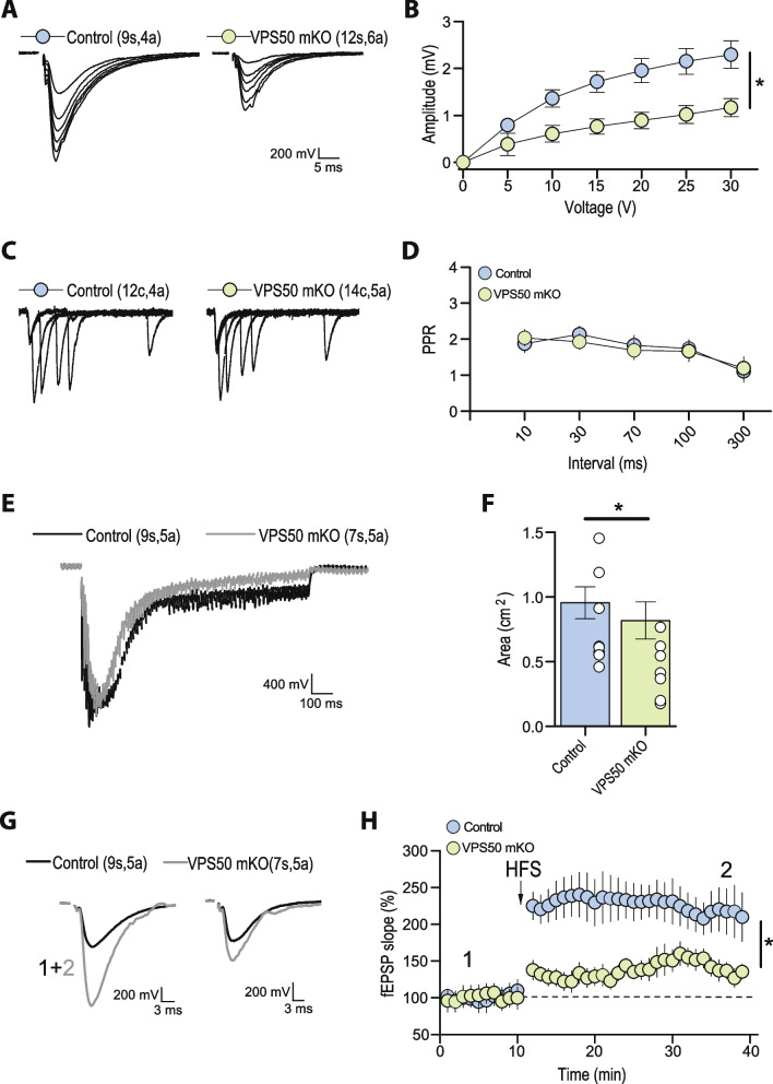
Fig. 6.
Hippocampal synaptic function and plasticity are impaired in VPS50 mKO mice. Electrophysiological recordings characterizing Schaffer collateral-to-CA1 synapses in acute hippocampal slices of control and VPS50 mKO animals. A Representative traces fEPSP of input–output curve elicited at different stimulus intensities. B Input–output curves reveal a strong reduction in the amplitude of fEPSC at all stimulus intensities tested. C Representative traces and D quantification of paired-pulse facilitation at different inter-stimulus intervals. E Representative synaptic responses and F quantification showing a strong synaptic depression in response to a single high-frequency stimulus train (100 pulses at 100 Hz) that likely reflect a reduce vesicular content in VPS50 mKO synapses compared to control synapses. G Representative traces before and after LTP induction elicited by four trains of high-frequency stimulation. Sample traces were taken at times indicated by numbers in summary plot. H Summary plot showing that the magnitude of LTP is reduced in VPS50 mKO animals. In each representative trace, the number of slices (s) or cells (c) and animals (a) are indicated in parenthesis. Two-samples Student t-test was used for statistical analysis; *p < 0.05. Summary data consist of mean ± SEM

