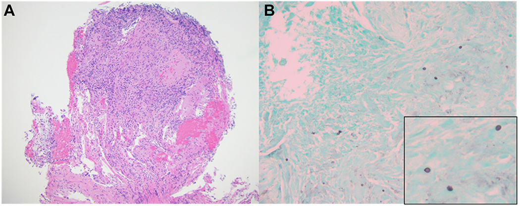
Figure 2 –

A, B, Photomicrographs showing granuloma with focal caseous necrosis (A) (Hematoxylineosin stain, original magnification ×100) and Gomori-methenamine silver staining of scattered yeast forms in the periphery of necrotic focus measuring approximately 5 μm in diameter (B) (Gomori-methenamine silver staining, original magnification ×200; inset, ×600 magnification).
