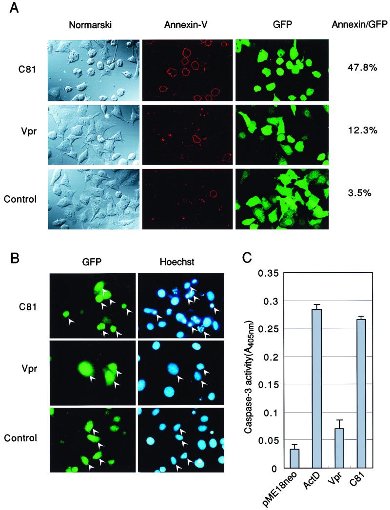
FIG. 2.
Expression of C81-induced apoptosis in HeLa cells. HeLa cells were transfected with pME18Neo that encoded Flag-tagged wild-type Vpr or Flag-tagged C81 or with the control pME18Neo-Flag together with (A and B) or without (C) the GFP expression vector pEGFP-N1. GFP was used as the reporter molecule for discrimination between transfected and untransfected cells. (A) Thirty-six hours after transfection, cells were stained with annexin V-biotin and streptavidin-PE for identification of apoptotic cells. The percentage of annexin V-positive cells relative to GFP-positive cells is indicated at the right. (B) HeLa cells were fixed in 1% formaldehyde and then in 70% ethanol and stained with Hoechst 33258 to monitor morphology. Apoptotic bodies (arrowheads) were revealed by fluorescence microscopy. (C) HeLa cells were harvested 36 h after transfection, and then caspase-3 activity was measured with a colorimetric kit. ActD, actinomycin D.

