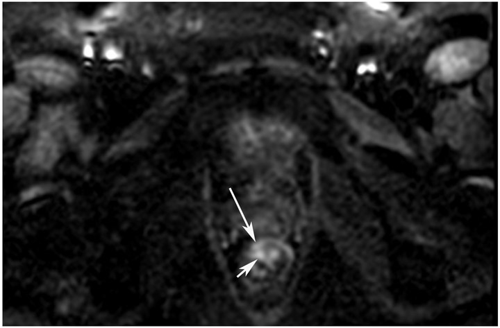
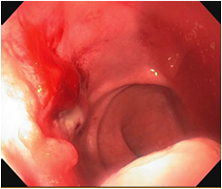
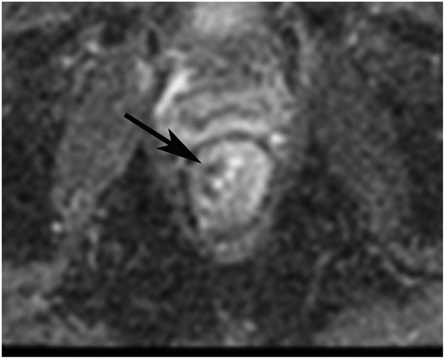
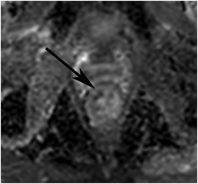
FIGURE 10:
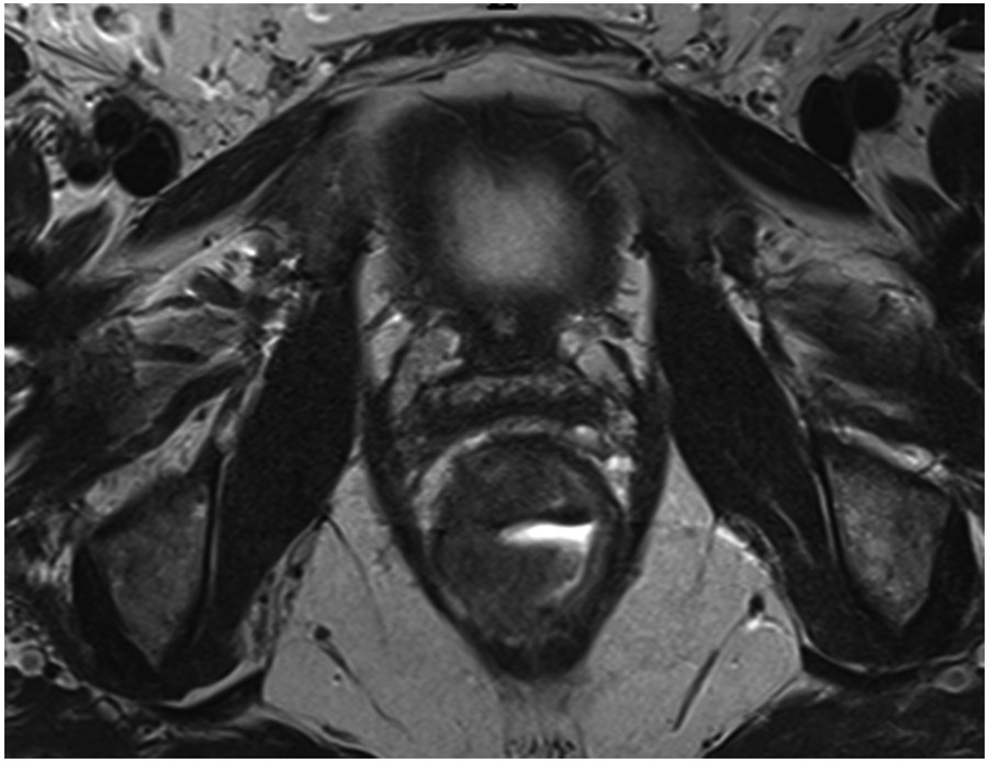
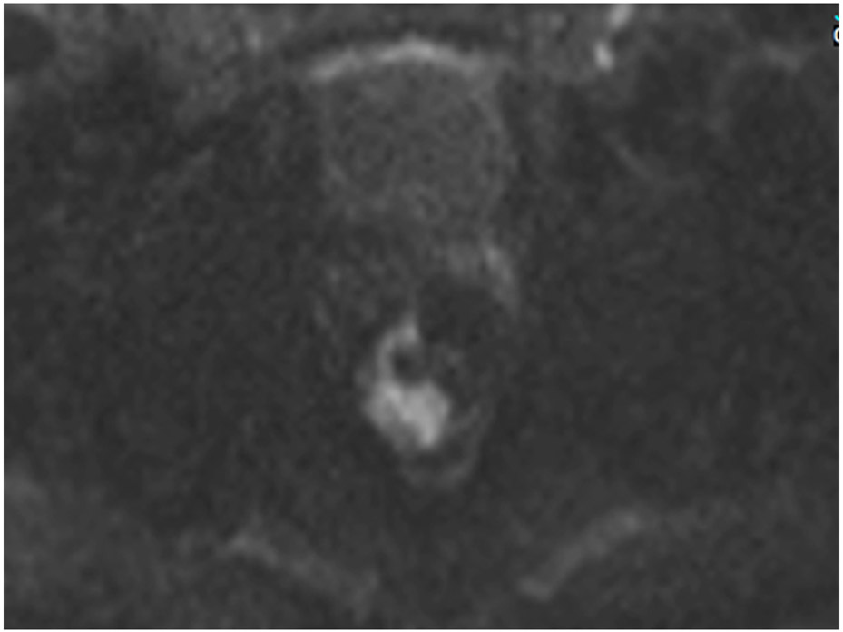
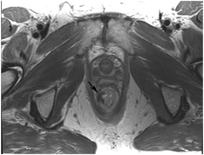
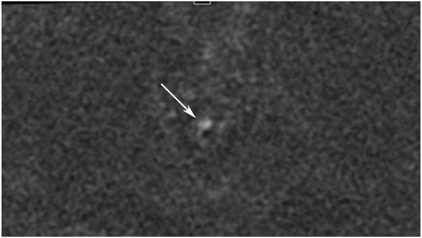
nCR 7 months after treatment. 61-year-old woman with T3N+ rectal cancer 2.6 cm from the anal verge underwent MRI at an external facility. (a) Straight axial 3-mm slice MRI through the tumor bed reveals a T2-intermediate signal partly circumferential tumor. (b) Also at baseline, DWI shows expected restriction of the tumor (high b-value). (c) Straight axial 5-mm slice T2WI at 10 months reveals a dark scar at tumor attachment site. (d) Matching b1500 FOCUS DWI. Note how the high signal is peripheral to the lumen and is in the wall (arrow). (e) Matching b800 image offered to show that even with the higher signal (T2-effects) seen in this lower b-value image, the pattern of more outer curvilinear peripheral signal in wall is distinguished from the inner mucosal pattern (short arrow). (f) Endoscopy shows regression of the tumor with persistent tumor nodules. (g–h) ADC maps for b1500 (g) and b800 (h) reveal a dark signal in the tumor bed (arrow) proving tumor rather than T2 shine-through. This patient did not continue to regress but rather the tumor increased, and she required surgery 3 months later (pT2N0).
TEACHING POINT: Nearly all the tumor is gone. There is only a T2 scar and a small focus of restriction. This is a good partial response, but tumor nodules on endoscopy at the same time prevented this from being declared “near complete response.” Pattern recognition and close comparison of the location of the tumor between matched bed positions on T2WI, DWI, and the ADC map are required to differentiate any remaining signal from the collapsed mucosa and from T2 shine-through.

