Figure 14:
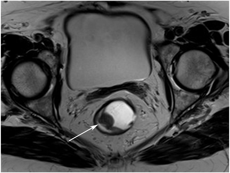
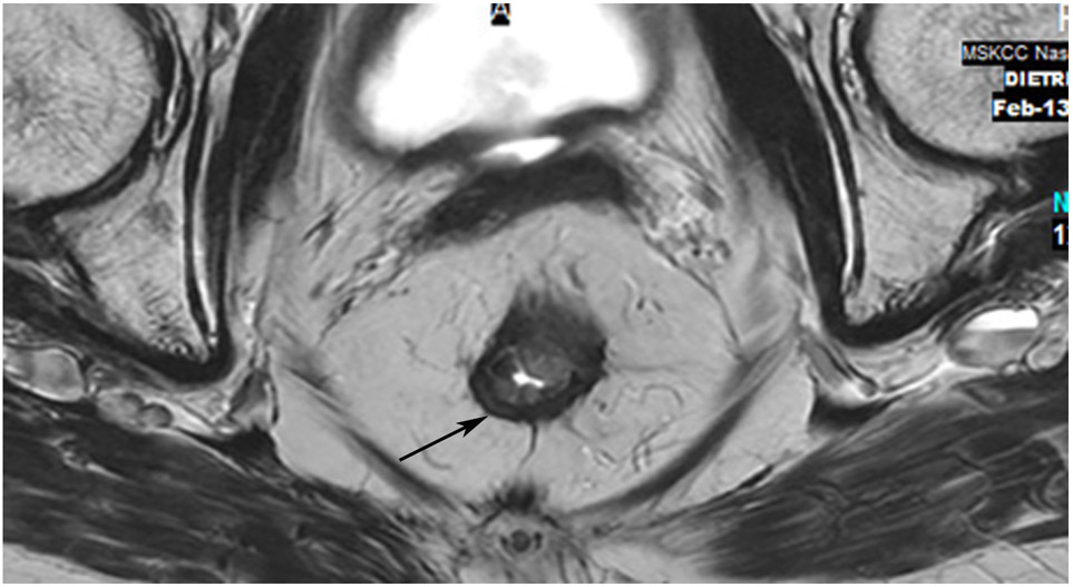
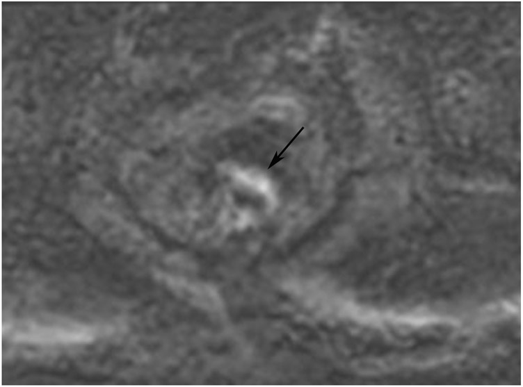
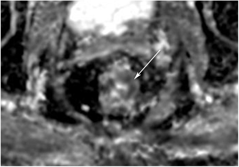
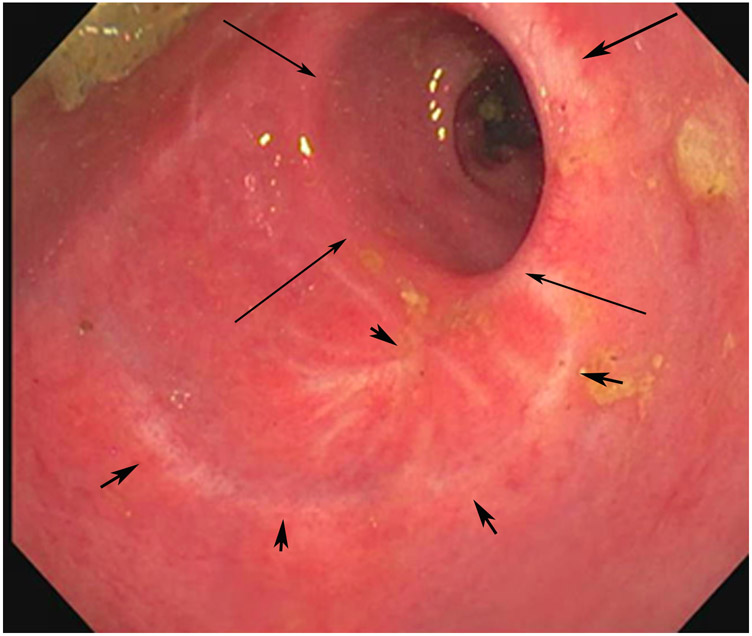
A 75-year-old woman presented for second-opinion post transanal excision (TAE) of a mass – cT1NxM0 adenocarcinoma with lymphovascular invasion and perineural invasion – with high-risk features but negative margins. (a) External facility pre-TAE 5-mm straight axial MRI slice shows a polypoid mass on fold (arrow). (b) 2 years after CRT (CRT was given in this case in light of the 10–20% risk of recurrence of T1 tumors and because a true cancer operation had not been performed), straight axial T2WI shows a scar in the tumor bed (arrow). (c) The interpreting radiologist trainee noted DWI restriction (arrow) OPPOSITE the tumor bed. Again, this should not be of any concern and is best ignored. Only the tumor bed on all slices should be at risk for regrowth. (d) ADC map shows that the DWI signal is true restriction, not shine-through (arrow). (e) Endoscopy indicates a well-healed TAE scar (small arrows) and stricture on the opposite wall (long arrows). It is hypothesized that stricture can cause restricted diffusion and that when known or present, interpretation caution is advised.
TEACHING POINT: DWI restriction away from the points of the initial tumor attachment and subsequent scar (here opposite wall!), should NOT represent tumor. Strictures are a special case and may cause false-positive DWI, possibly due to stricture-induced restricted proton motion compared with that of the normal wall. It is too early to state this with confidence, but if a stricture is noted at MRI or endoscopy, interpretive caution is advised.

