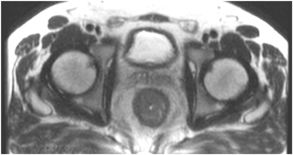
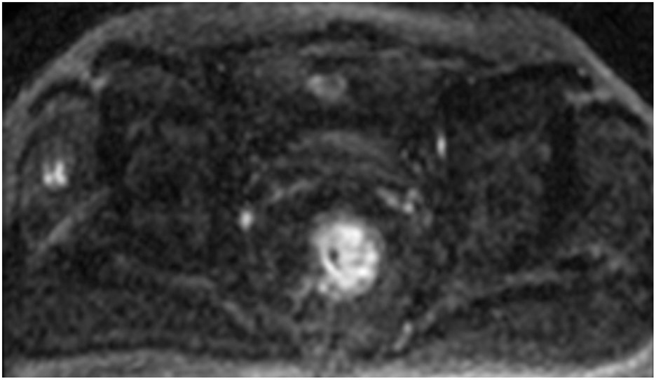
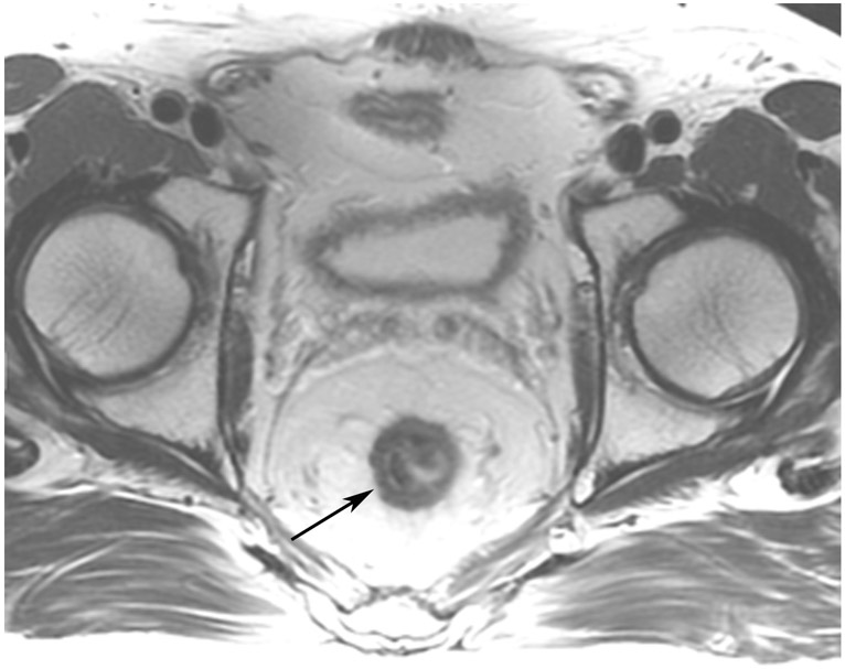
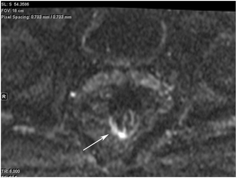
Figure 16:
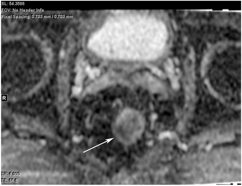
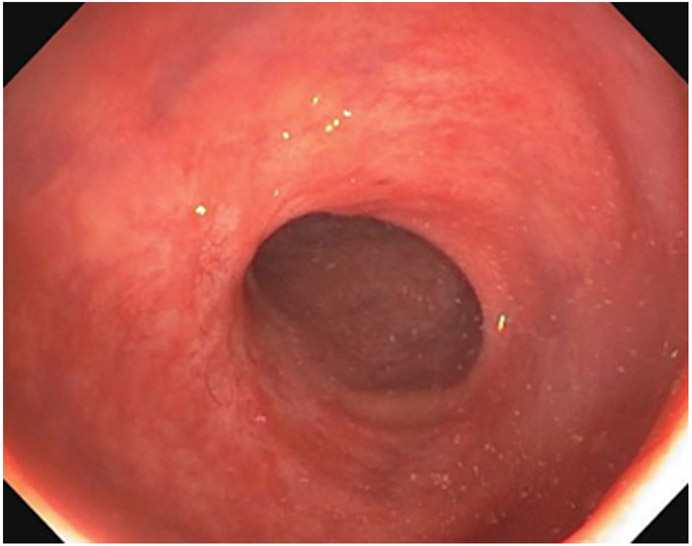
A 44-year-old woman with a rectal mass underwent MRI at an external facility. (a) 5-mm straight axial T2WI slice shows a circumferential tumor. (b) Matching 6-mm axial b800 DWI slice reveals circumferential diffusion restriction. (c) 1.5 years after TNT, surveillance MRI shows a scar (arrow). (d, e) Matching DWI shows a high signal along with diffusion restriction on the ADC map (arrows). (f) Endoscopy, however, reveals no tumor. 6 months later, the patient is still free of disease.
TEACHING POINT: Most mismatched findings of restricted signal on DWI and normal endoscopy (80% per one series) will prove to be false-positive DWI findings, indicating that endoscopy more accurately finds cCR. There are myriad causes of false positives, including inflammation, stricture, artifact, adenoma without cancer and/or hyperplastic mucosa, perceptive error, and interpretive error (looking at the wrong area).

