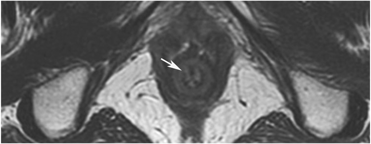
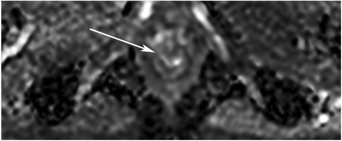
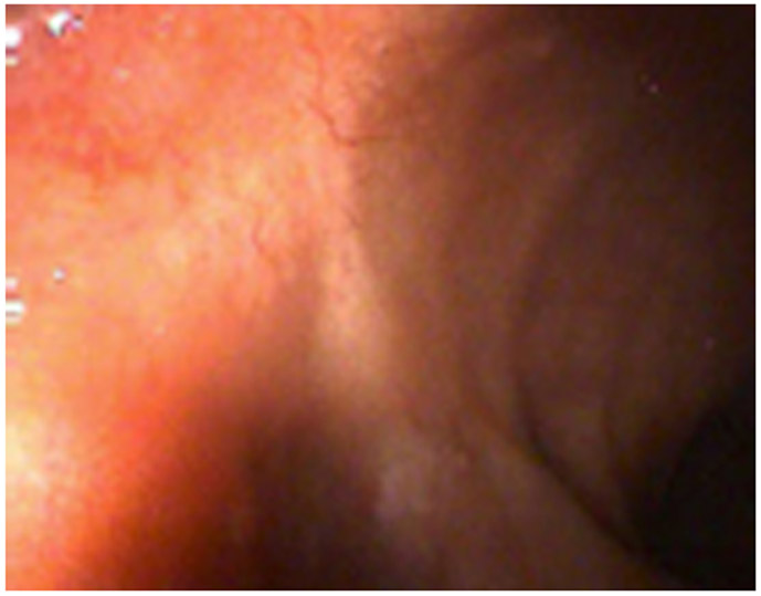
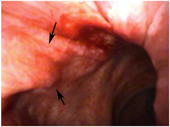
Figure 18:
44-year-old man with a treated rectal mass underwent surveillance scans 4 months apart after the completion of TNT. (a) 5-mm straight axial T2WI slice reveals a collapsed mucosa with bright T2 signal (arrow) (one slice 3 mm below the scar is not shown). (b) Matching axial b800 DWI slice reveals T2 shine-through in the same pattern (arrow), i.e., tri-radiate “Mercedes-Benz sign,” as that of the T2 bright collapsed mucosa. (c) ADC map confirms T2 shine-through (arrow). (d) Endoscopy is normal. (e) 4 months later, axial T2WI at the same level shows the same tri-radiate pattern (arrowheads). (f) DWI also shows a similar tri-radiate pattern but with a subtle difference on direct comparison, i.e., the left anterior portion of collapsed mucosa is globular (arrow), different from the classic Mercedes-Benz sign (also see schematic). (g) ADC map shows a new dark signal at the point of the globular configuration (arrow), indicating diffusion restriction suspicious for tumor. (h) Endoscopy shows new mucosal coarsening and nodularity (arrows), suspicious for regrowth. The patient underwent brachytherapy but the tumor regrew, requiring low anterior resection and then abdominoperineal resection; the tumor then metastasized to the inguinal and retroperitoneal nodes and resulted in lung metastases. 8 years from diagnosis, the patient is still alive and undergoing chemotherapy.
TEACHING POINT: The collapsed mucosa may look tri-radiate (Mercedes Benz sign) or have more extensions (e.g., quadra-radiate, penta-radiate) and its recognition can help with discerning subtle changes.


