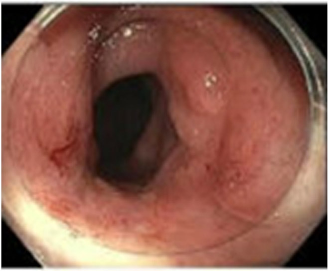
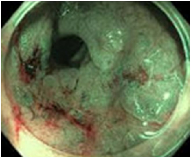
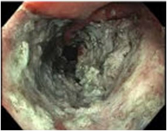
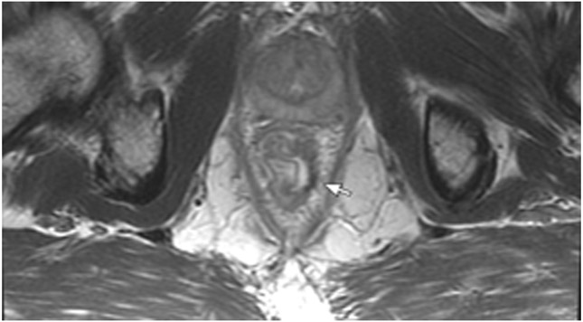
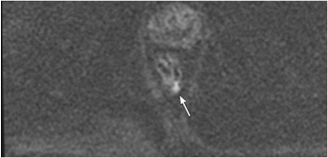
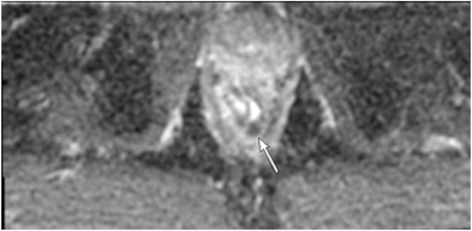
Figure 23:
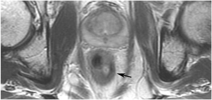
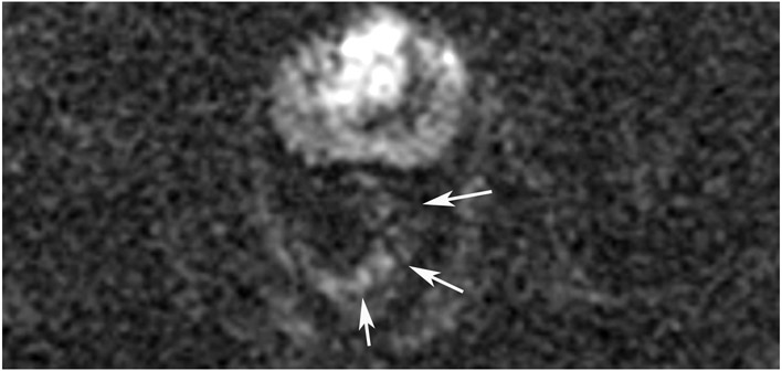
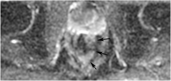
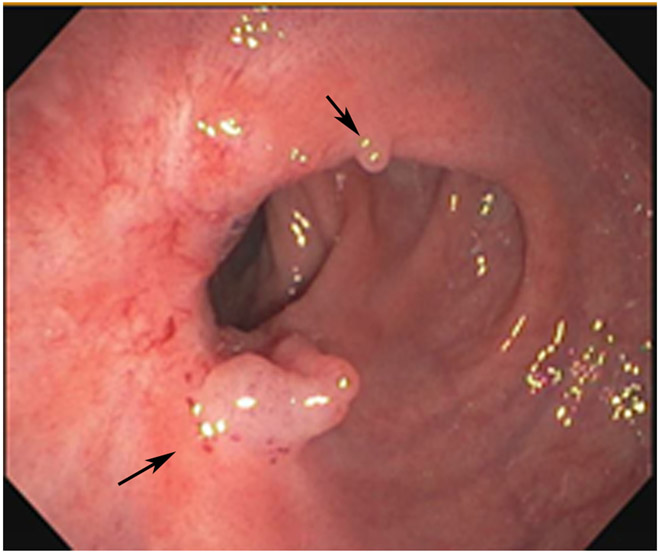
63-year-old man with primary rectal cancer 5.2 cm from the anal verge underwent follow-up imaging post treatment. (a) Post-TNT scan at 3 months. Axial T2WI shows treated tumor bed with scar (arrow). (b, c) Axial b1500 DWI FOCUS (b) and the ADC map at the same level reveals T2 shine-through when DWI is paired with ADC (c) (arrows). There is no true restriction. (d) Endoscopy, however, reveals “residual adenoma or redundant mucosa”, (arrows). The patient was referred for ESD. (e–g) ESD images: e) white light, f) narrow band imaging, and g) post-ESD image (images courtesy, Dr. Makoto Nishimura). Pathology revealed pT1Nx tumor with a positive margin. (h–j) 1 month later, follow-up axial T2WI (h) reveals a similar appearing scar (arrow), and restricted diffusion (arrows; i; DWI b1500, j; ADC map). This patient required abdominoperineal resection (APR) and was pT3N0. 2 years after APR, the patient is disease-free.
TEACHING POINT: Residual, often pre-existent adenoma, not responsive to treatment, may be left over and may not be detected by MRI. It may or may not show DWI restriction and it may or may not have tumor in it. It is a limitation of MRI requiring more study.

